18- BODY FLUIDS AND CIRCULATION
CHAPTER NO.18 BODY FLUIDS AND CIRCULATION
A178
INTRODUCTION:As
we know, all living organisms are made up of cells .All living cells have to be
provided with nutrients, Oxygen and other essential substances .Also the waste
and harmful substances produced, have to be removed continuously for the
healthy functioning of tissues. Therefore, it is
essential to have efficient
mechanisms for the movement of these substances to
the cells and from the cells.
Blood is the most commonly used body fluid by most
of the higher organisms including humans for this purpose.
Blood: Blood is a special connective tissue
consisting of a fluid matrix called
plasma and formed elements.
PLASMA:It is Straw coloured, viscous fluid
constituting nearly 55% of the blood.
90 -92 percent of plasma is water and protein
contributes 6 - 8 percent of it.
Three types of protein are present in plasma:
1. EERE - Helps in blood clotting or coagulation.
2. TE - !nvoived in the defence mechanism of the
body.
3. EY - Helps in maintaining osmotic balance.
Plasma also contains small amounts of minerals like
Nat+,Ca++,Mb++, HCO3-,Cl-
etc.Factors for blood coagulation are also present
in plasma in an inactive form.
Plasma without clotting factors is called Serum.
FORMED ELEMENTS:
There are three types of formed elements:
1. Red blood cells or Erythrocytes
2. White blood cells or Leukocytes
3. Platelets or Thrombocytes
1. ERYTHROCYTES
or RBC are the most abundant of all the cells in blood
(5 - 5.5 per cubic mm).Itis formed in red bone
marrow in adults.RBCs are devoid of nucleus and are biconcave in shape.
They have a red coloured ,iron containing complex
proteins called haemoglobin which give it red colour.Haemoglobin plays a
significant role in transport of respiratory gases.Life span of RBC is 120 days
after which they are destroyed in spleen which is called the graveyard of RBC.

2. LEUCOCYTES
or WBCs They are colorless due to absence of haemoglobin They are nucleated and
are lesser in number(6000-8000 per cubic mm)Leukocytes are generally short
lived Leukocytes are of 2 types
A. Granulocytes
B. Agranulocytes
A.GRANULOCYTES: These contain granules in the cytoplasm
and are of
three types-
1. Eosinophils(2-3%) resist infection and are
associated with allergic reaction
2. Basophils(0.5-1%) secrete histamine, serotonin
and heparin.and are involved in inflammatory reaction
3. Neutrophils(60-65%) are phagocytic cells which
destroy foreign organisms entering the body.
B. AGRANULOCYTES: They do not contain granules in
the cytoplasm. These are of two types-
1. Monocytes(6-8%) are phagocytic cells which
destroy foreign organisms
entering the body.
2. Lymphocytes (20-25%) are of 2 major types- B
Lymphocytes and T Lymphocytes.Both B and T Lymphocytes are responsible for
immune response of the body.
3. PLATELETS
Also called Thrombocytes.
These are formed in the bone marrow from the cell
fragments of megakaryocytes
Blood contains 1,50,000 -3,50,000 platelets per
cubic mm Platelets help in coagulation or clotting of blood Reduction in their
number leeds to clotting disorders which will lead to excessive loss of blood
from the body.
LET US KNOW WHAT WE HAVE LEARNT!
1. Multiple Choice Questions:
|. Which one of
the following types of cells lacks a nucleus?
(a)RBC
(b)Neutrophil
(c)Eosinophils
(d)Monocytes
ll. Cells
involved in inflammatory reaction are:
(a)Basophil
(b)Neutrophil
(c)Eosinophils
(d)Lymphocytes
Ill. Mark , among
the following a cell which exhibit phagocytic activity:
(a)Neutrophil
(b)Basophil
(c)Eosinophils
(d)RBCs
IV. Graveyard of
RBCs is
(a)Liver
(b)spleen
(c)kidney
(d)Thymus
V. Which
Leukocyte releases Heparin and Histamine in the blood
(a)Neutrophil
(b)Basophil
(c)Eosinophils
(d)Monocytes
2. True/False:
|. Life span of RBCs is 120 days.
Il. Leukocytes help in blood coagulation.
Ill. Monocytes and neutrophils are phagocytic cells.
3. Fill in the Blanks:
|. Plasmawithout__is called serum.
ll. ~~ and Monocytes are phagocytic cells.
lll. Eosinophils are associated with
————_—sreactions.
ANSWER KEY: PART A
1. Multiple choice questions:
Ans 1:(a)RBC.
RBC is a nucleated cell.
Ans 2:(a)Basophils
Basophils are involved in inflammatory reactions.
Ans 3:(a)Neutrophil
Neutrophils destroy the foreign organism entering
the body.
Ans 4:(b)spleen
RBCs are destroyed in spleen.
Ans 5:(b)Basophil
Basophil releases Heparin and Histamine in blood.
2. True/False:
|. True
ll. False-Platelets help in blood coagulation
Ii. True
3. Fill in the Blanks:
|. clotting factors
li. Neutrophils
Ii. = Allergic
1. Why erythrocytes are called Red Blood Cells?
2. What is the importance of Plasma proteins?
3. Why do we consider blood as a connective tissue?
1. Name the components of Formed Elements in the
blood and mention one major function of each of them?
A179
INTRODUCTION:In the last
assignment we studied blood and its components. As we know, the blood of human
beings differs in certain aspects though it appears to be similar.Various types
of blood grouping have been done. Two very important and common types of blood
group systems are:
1. ABO Blood groups
2. Rh blood group system
ABO Blood groups: It is based on the presence or
absence of two surface
antigens on the RBCs namely Similarly plasma of
different individuals
contains two natural antibodies. The distribution of
antigens and antibodies in four groups of blood are Blood group A has A antigen
and antibody Blood group B has B-antigen and bantibody a Blood group AB has
both A and B antigens but no antibody
Blood group O has no antigens but with both
antibodies a & b During the blood transfusion, any blood cannot be used.
The blood of a donor has to be carefully matched with the blood of the
recipient before any blood transfusion to avoid severe problems. The donor's
compatibility is shown in the following table.
UNIVERSAL DONOR:
The person with O blood group is called Universal donor as
it has no antigen and can donate its blood to a
person with any blood group.
UNIVERSAL RECIPIENT:
The person with AB blood group is called Universal
recipient as it has no antibody in their plasma so
can receive blood from any blood
group.
RH BLOOD GROUP SYSTEM:
Rh factor is an antigen protein present on the surface of RBCs.It was first
discovered on the plasma membrane of RBCs of Rhesus monkey, so is called Rh
factor. Later it was found in about 94% of human beings and was called Rh
positive (Rh+). The person with no Rh factor is called Rh negative (Rh-).
An Rh -ve person, if exposed to Rh +ve blood, will
form specific antibodies against the Rh antigens .Therefore the Rh group should
also be matched before
transfusions.A special case of Rh incompatibility
has been observed between the Rh-ve blood of pregnant mother with Rh+ve blood
of the fetus. Rh antigen of the foetus do not get exposed to the Rh-ve blood of
the mother in the first pregnancy as the two bloods are well separated by
placenta. However during the delivery of the first child, there is possibility
of exposure of maternal blood to small amounts of Rh+ blood from the foetus. In
such case mother starts preparing antibodies against Rh antigens in her blood.
In her second pregnancy Rh antibodies from the mother
(Rh-ve) can leak into blood of the foetus (Rh +ve)
and destroy the foetal RBCs.
This could be fatal to the foetus or could cause
severe anemia and jaundice to the
baby. This condition is called erythroblastosis
foetalis .This can be avoided by administering anti -Rh antibodies to the
mother immediately after the delivery of the first child.
COAGULATION/CLOTTING OF BLOOD
DEFINITION:
The property of blood to change from fluid to gel state when coming
in contact with air in response to an injury or
trauma is called blood clotting.
AIM: To prevent excessive loss of blood from an
injury, so is a natural defensive
mechanism.
MECHANISM:
When there is an injury in the body, it stimulates the platelets in the
blood to release Thrombokinase or Prothrombinase
which activate the
mechanism of coagulation. The injured tissue can also
initiate blood coagulation by releasing Thromboplastin which gets converted
into Thrombokinase /
Prothrombinase. In the presence of Calcium ion,
Thrombokinase activates prothrombin and from Thrombin. Prothrombin is formed by
the liver in the presence of vitamin K. Thus Calcium ion and vitamin K play
important roles in blood clotting.
Thrombin acts on fibrinogen, present in plasma and
converts it into fibrin. Fibrin
threads polymerize and forms a mesh work which traps
blood cells, platelets etc. to
form a clot. So the process is called blood
clotting.

LET US KNOW WHAT WE HAVE LEARNT!
PART A: VERY SHORT ANSWER TYPE
QUESTIONS:
1. Multiple Choice Questions:
|. Vitamin which
is essential for blood clotting is
(a)Vitamin A
(b)Vitamin B
(c)Vitamin C
(d)Vitamin K
ll. The most
important mineral for blood coagulation is
(a)Calcium
(b)Magnesium
(c)Sodium
(d)lron
lil. | Which
blood group is called Universal recipient
(a)Blood Group A
(b)Blood Group B
(c)Blood Group AB
(d)Blood Group O
IV. Which blood
group is called a Universal donor?
(a) Blood Group A
(b) Blood Group B
(c) Blood Group AB
(d) Blood Group O
V. Which of the
following are involved in blood clotting at the injury?
(a) Platelets
(b) RBCs
(c) Neutrophils
(d) Monocytes
2. True/False:
|. Aperson with B blood group cannot donate blood to
a person of A
blood group.
Il. | Blood group is designated on the basis of
presence of antibodies in the
blood plasma.
Ill. Aperson with AB blood group is a universal
recipient.
3. Fill in the Blanks
l.
w.............40N plays an important role in the blood clotting.
ll. | Rh factor was first discovered on the plasma
membrane of RBCs of....
lll. Aperson of O blood group has ........and
.........antibodies in his blood plasma.
ANSWER KEY: PART-A
1. MULTIPLE CHOICE QUESTIONS:
Ans 1: (d)vitamin K.
Vitamin K is required for the formation of
Prothrombin in the liver.
Ans 2: (a) Calcium
Calcium ion is required for the formation of
thrombin from prothrombin
Ans 3: (c) Blood group AB
Blood group AB has no antibody in their plasma so
can receive blood from any blood group.
Ans 4: (d) Blood group O
Blood group O has no antigen and can donate blood to
any person.
Ans 5: (a) Platelets
At the site of injury Platelets release
thrombokinase or Prothrombinase which activate the mechanism of coagulation.
2. TRUE/FALSE:
|. True
|. False-Blood group is designed on the basis of
presence of antigens on
the surface of RBCs.
Ii. True
3. FILL IN THE BLANKS:
|. Calcium
ll. Rhesus Monkey
Il. Anti A and Anti B
PART B: SHORT ANSWER TYPE QUESTION:
1. What is Erythroblastosis foetalis?
2. Person with blood group O is called Universal
donor. Why?
3. Thrombocytes are essential for coagulation of
blood. Comment.
PART C: LONG ANSWER TYPE QUESTION:
1. Explain different types of blood groups and donor
compatibility by making
a table.
A180
INTRODUCTION:It
is a clear to pale white fluid derived from tissue fluid which
circulate throughout the lymphatic system.
The main function of lymph is to protect the body
from harmful germs.Lymph is also called the middleman of the body.
COMPOSITION OF LYMPH
1. Lymph plasma
2. Lymph Corpuscles
3. Lymphoid Organs
LYMPH PLASMA
It consists of less calcium, few blood proteins which includes albumin :
globulin to be 1:5, fibrinogen content is low along with that of phosphorous.
Globulin proteins which are actual antibodies are
found in lymph plasma.
LYMPH CORPUSCLES
These are leucocytes (White Blood Cells) and amoeboid cells. Erythrocytes and
blood platelets are absent.
LYMPHOID ORGANS
Numerous lymph nodes are present deep inside the body.These lymph nodes are
connected to lymphatic vessels which circulate the lymph throughout the
body.The lymph gets filtered at the lymph nodes.The spleen, tonsils, adenoids
and thymus are also the lymphoid organs.
The spleen is considered as the largest lymphatic
organ in the system. An
elaborate network of vessels is called the Lymphatic
System.Spleen maintains mature new lymphocytes and also synthesizes
antibodies.Lymph and tissue fluid both have blood clotting property.

OTHER COMPONENTS OF LYMPH
1. Carbohydrates
2. Lymphocytes
3. Water
4. Urea
5. Chlorides
6. Enzymes
7. Very low amount of fat
8. Proteins
9. Non Protein Substances.
FUNCTIONS OF LYMPH
Lymph performs many important functions. Few major functions of
lymph are mentioned below:
1. Itkeeps the body cells moist.
2. It transports oxygen, hormones and nutrients to
different parts of the body.
3. Itremoves metabolic waste from the cells.
4. It transports antibodies and lymphocytes to the
blood.
5. It prevents entry of microbes inside the lymph
nodes.
LET US KNOW WHAT WE HAVE LEARNT!
PART: A VERY SHORT ANSWER TYPE
QUESTIONS:
(A) MULTIPLE CHOICE TYPE QUESTIONS
1. What is the
fluid that flows in the lymphatic system?
a) Blood
b) Lymph
c) Urine
d) None of the above
2. One of the
main component of lymph is:
a) Red Blood Cells
b) Bile
c) White Blood Cells
d) None of the above
3. What kind of
cells do lymph nodes produce?
a) Lymphocytes
b) Red Blood Cells
c) Nerve Cells
d) None of the above
4. Which of these
is a part of the the lymphatic system?
a) Spleen
b) Thymus
c) Tonsils
d) All of these
5. Largest
lymphatic organ is
a) Spleen
b) Thymus
c) Tonsils
d) None of these
(B) FILL. IN THE BLANKS:
(a)............... proteins which are actual
antibodies are found inlymph
plasma.
(b)..............., tonsils and thymus are also the
lymphoid organs.
(C )................. removes metabolic waste from
the cells.
(C) TRUE /FALSE
(a) Lymph prevents entry of microbes inside the
lymph nodes.
(b) Blood is the fluid that flows in the lymphatic
system.
(c ) Lymph keeps the body cells moist.
ANSWER KEY: PART-A
(A) MULTIPLE CHOICE TYPE QUESTIONS :
1. (b) Lymph (Lymph is the fluid which flows in the
Lymphatic
system).
2. (c) White Blood Cells (The main component of
lymph is
White blood cells).
3. (a) Lymphocytes (Lymph Nodes produce
lymphocytes).
4. (d) All of these (Spleen, Tonsils, thymus are the
parts
of Lymphatic System).
5. (a) Spleen (Spleen is the largest Lymphatic
Organ).
(B) FILL IN THE BLANKS:
(a) Globulin Proteins
(b) Spleen
(c) Lymph
(C) TRUE /FALSE
(a) True
(b) False (Lymph is fluid that flows in the
lymphatic system).
(c ) True
PART: B SHORT ANSWER TYPE QUESTIONS:
1. Name the Lymphoid Organs.
2. Write the composition of Fluid.
3. Name the Lymph corpuscles.
PART: C - LONG ANSWER TYPE QUESTION:
1. What are the components and functions of Lymph?
A181
INTRODUCTION:HEART:The
mesodermally derived organ is situated in the thoracic cavity, in between two
lungs slightly tilted to the left. It has the size of clenched fist. It is
protected by a double walled membranous bag pericardium enclosing the
pericardial fluid.
STRUCTURE OF HUMAN HEART:Our
heart has four chambers, two relatively small upper chambers called atria and
two larger lower chambers called ventricles.A thin muscular wall called the
inter-atrial septum, separates the right and left atria whereas a thick walled
inter ventricular septum, separates the left and the right ventricles.The atrium
and the ventricles of the same side are also separated by a thick
fibrous tissue called the atrio- entricular septum.However, each of these septa is provided with an opening through which the two chambers of the same side are connected.

The opening between the right atrium and the right ventricle is guarded by a valve formed of three muscular flaps. The tricuspid valve, whereas bicuspid or mitral valve guards the opening between the left atrium and the left ventricle.

The
opening of the right and left ventricles into the pulmonary artery and the
aorta
is provided with semilunar valves.The valves in
heart allow flow of blood in one direction.The entire heart is made up of
cardiac muscles.A patch of tissues called sinoatrial node is present in right
atrium.
Another mass called atrio ventricular node is
present in left comer of right atrium.
A bundle of nodal fibres which passes through the
atrio -ventricular septa to inter ventricular septum divides into right and
left bundle are called PURKINJE FIBRES
or BUNDLE OF HIS. These can generate the maximum of
action potentials. 70-75
min. and responsible for maintaining rhythmic
contractile activity of heart.
LET US KNOW WHAT WE HAVE LEARNT!!
PART: A VERY SHORT ANSWER TYPE
QUESTIONS:
a) MCQs:
1. How many
chambers human heart has?
a) 1
b)2
c)3
d)4
2. The opening
between the right atrium and right ventricle is guarded by
a) Tri cuspid valve
b) bicuspid valve
c) none of above
d) both
3. Where is sino
atrial node situated?
a) Left auricle
b) right auricle
c) right ventricle
d) left ventricle
4. Our heart
normally beats:
a) 70-75/ min
b) 80-85/min
c)100-120/min
d) none of the above
5. Where is
purkinje fibres situated?
a) Auricles
b)ventricles
c)atrio ventricular septum
d) none of these
b) True/False:
1. Heart is protected by a single membrane.
2. Our heart has two chambers.
3. The valves in the heart allows the flow of blood
in one direction
c) Eill ups:
1. Mass of the tissues is seen in the lower left
corner of right atrium is
2. Our heart beats normally....... times in a
minutue.
Answer key: Part—A
b) MCQs:
1. d Chambers in heart=4
2. a Tricuspid valve
3. b Right auricle
4. a 70-75/ min
5. c atrio
ventricular septum
d) True/False:
1) False: Heart is protected by a double membrane.
2) False: Our heart has four chambers.
3) True
e) Fill ups:
1) atrio ventricular node
2) 70- 75/min
PART: B_ SHORT ANSWER TYPE QUESTIONS:
Q1. Sino-atrial is called the pacemaker of our heart
?
Q2. What are semilunar valves?
Q3. Where is Purkinje fibres situated?
Q4. How action potential is generated in heart
PART: C_ LONG ANSWER TYPE QUESTIONS:
Q1. Explain internal structure of human heart with
well labelled diagram.
A182
INTRODUCTION:When
multicellular organisms need a system to transport oxygen and
nutrients throughout the body, that system is known
as the Circulatory system. In living organisms, the circulatory system has two
pathways. These two circulatory pathways are:
Open type
Closed type
Open circulatory system:It is the one in which blood
pumped by the heart passes through large vessels into open spaces or body
cavities called Sinuses. Example:
arthropods and molluscs.

Open Circulatory System is
the system that is primarily found in invertebrates. In open circulatory
system, the blood is not confined within
blood vessels. The blood bathes the organs and
tissues directly. Because of
this, the blood and the interstitial fluid have no
distinction. Thus, the term
haemolymph is used. The haemolymph is pumped by the
heart is pumped by
the heart through the vessels into the sinuses and
then to the surrounding
cells where exchange of materials occur. The
haemolymph is taken back to
the heart through ostia that close via the valves
during the heart contraction.
The haemolymph is composed of fluid plasma (containing water, organic compounds, and inorganic salts such as Na*, Cl, Mg**, and Ca?*). There are also free floating cells called haemocytes that are involved in the immune function.
Closed circulatory system
In this, the blood pumped by the heart is always
circulated through a closed
network of blood vessels. Example: Annelids and chordates. In closed Circulatory System, the presence of vessels conducts blood throughout the body. Blood flows inside the body through vessels such as arteries, veins and capillaries.

Closed type circulatory system is more advantageous as the flow of fluid can be more precisely regulated.

Human
circulatory system
Human beings follow a closed circulatory system
which means that the blood
is always enclosed in vessels and the heart while circulating
throughout the
body. Travelling through arteries and veins, blood
carries important
molecules throughout the body and is always
confined.Advantage of Closed Circulatory System Compared to the open
circulatory system, the closed circulatory system
operates with much higher blood pressure. It is said
to be more efficient
considering that it uses much less blood for even
faster and higher ievels of
distribution.
LET US KNOW
WHAT WE HAVE LEARNT!!
PART: A VERY
SHORT ANSWER TYPE QUESTIONS
1.Which of the
following organism has an open circulatory system?
(a) Humans
(b) Birds
(c) Dogs
(d)Grasshoppers
2.What is the
major differance between and open and closed circulatory system?
(a) Function
(b) Material transported
(c) Color of the blood
(d) Structure and design of vessels
3.An open
circulatory system describes?
(a) A circulatory system that is open to the
environment.
(b) A circulatory system in which blood and
interstitial fluid are allowed
to mix.
(c) The circulatory system found in humans.
(d) A circulatory system that has two loops: one for
oxygenated blood,
one for waste products.
4.What is the
purpose of the circulatory system?
(a) Digest food
(b) Transmit feelings
(c) Transport blood and nutrients
(d) Produce proteins
5.How many types
of circulatory pathways are present?
(a) 1
(b) 2
(c) 3
(d) 4
(2)TRUE OR FALSE
1. In open type circulatory system, the blood is
pumped out of the heart
and the cells and tissues are directly bathed in it.
2. Chordates have open type circulatory system.
3. Open type circulatory system is more efficient
than closed type
circulatory system.
(3)FILL IN THE BLANKS
1. In open type circulatory system, since blood and
interstitial fluids
have no distinction, term is used for that fluid.
2. Annelids and chordates have type of circulatory
system.
ANSWER KEY:PART-A
(MCQ)
1. (d) Grasshoppers
2. (d) Structure and design of vessels
3. (b) A circulatory system in which blood and
interstitial fluid are
allowed to mix.
4. (c) Transport blood and nutrients
5. (b) 2
(2)TRUE OR FALSE
1.True
2.False Chordates have closed type circulatory
system.
3.False Open
type circulatory system is less efficient than closed type
circulatory system. Closed circulatory system is
said to be more efficient considering that it uses much less blood for even
faster and higher levels of distribution.
(3)FILL IN THE BLANKS
1. Haemolymph
2. Closed
PART: B SHORT ANSWER TYPE QUESTIONS:
1. What do you mean by circulatory system?
2. Name the two types of circulatory pathways found
in animals.
3. What is the Advantage of Closed Circulatory
System?
PART: C LONG ANSWER TYPE QUESTIONS:
1. What is a circulatory pathway? Briefly describe
the types of circulatory
pathways with the help of well labelled diagram.
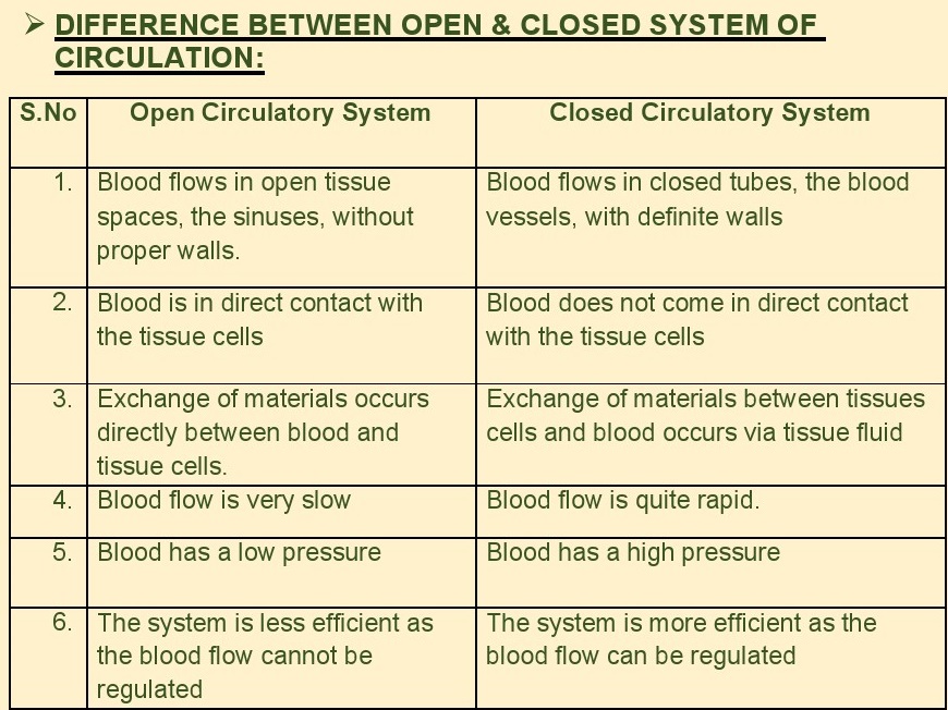
2. Differentiate between open and closed type circulatory
system ina
tabulated form.
A183
INTRODUCTION:Higher
vertebrates such as amphibians, reptiles, birds and mammals have arterio-venous
heart because both oxygenated (arterial) blood and
deoxygenated (venous) blood comes to it. Amphibians
have three chambered heart- two auricles and one ventricle. Reptiles have
incompletely four chambered heart. In crocodiles
(reptiles), birds and
mammals heart is four chambered — two auricles and
two ventricles. During
single cardiac cycle, blood goes twice in the heart
which is known as double circulation. In amphibians and reptiles (except
crocodile) there is mixing of oxygenated and deoxygenated blood in ventricle.
So, it is incomplete double circulation in these animals. Whereas perfect
separation of arterial and venous blood in birds and mammals, have ensured
complete double circulation. In double circulation, part of blood goes to lungs
for oxygenation. The oxygenated blood comes back to heart for pumping into
rest of the body. The two types of circulation are
called pulmonary
circulation and systemic circulation respectively.
1. PULMONARY CIRCULATION
- it is circulation of blood between heart and lungs. In Pulmonary circulation
pathway, the deoxygenated blood is pumped out from the right atrium into the
right ventricle as the tricuspid valve opens. This blood is then carried away
from the heart by pulmonary arteries to the lungs. In the lungs, the gaseous
exchange occurs (as CO2is released and O2 is picked up) and deoxygenated blood
is purified and
oxygenated. After that, the pulmonary veins bring
back the oxygenated blood to the left atrium of the heart. This blood then
enters the left ventricle from where it can be circulated throughout the body
parts.

2. SYSTEMIC CIRCULATION: Here the blood
circulates between heart and all parts of the body except lungs. In systemic
circulation, the heart supplies oxygenated blood from left atrium to the left
ventricle via bicuspid valve. From left ventricle, blood enters aorta. From
there, the blood travels through large arteries finish up in the capillary
network. In the
process, the blood supplies necessary oxygen and
other nutrients to tissues and collects the residual substances and carbon
dioxide from there and returns deoxygenated blood into the right atrium and
ventricle of the heart. Both pathways of blood circulation work together. Since
blood has to reach every tissue and cell, the blood pressure in systemic
circulation is
more than pulmonary circulation. ine separation of
oxygenated and deoxygenated blood in double circulation allows for an efficient
supply of oxygen to the body cells and delivers a greater blood flow rate.

PORTAL SYSTEM
Normally veins after collection of blood carry it to the heart. The vein which
after collection of blood carries and distributes it to another organ instead
of heart is called portal system. Thus, portal veins act both as collecting and
distributing vessels.
HEPATIC PORTAL SYSTEM:A
unique vascular connection exists between the digestive tract and liver called
hepatic portal system. Hepatic portal veins carries blood from intestine to the
liver before it is delivered to the systemic circulation. It helps in transport
and assimilation of digested food in liver.A special coronary system of blood
vessels is present in our body exclusively for the circulation of blood to and
from the cardiac muscles.

LET US KNOW WHAT WE HAVE LEARNT!!
PART: A VERY SHORT ANSWER TYPE
QUESTIONS:
1. Which of the
following statements is incorrect about double circulation?
(a) It occurs only in mammals.
(b) It occurs in all vertebrates.
(c) It is composed of systemic circulation.
(d) It is composed of pulmonary circulation.
2. How many
chambers are present in the heart of crocodiles?
(a) Two
(b) Three
(c) Four
(d) Five
3. Which of the
following organisms show incomplete double circulation?
(a) Birds
(b) Reptiles
(c) Mammals
(d) Crocodiles
4. Systemic
circulation in the cardiac system refers to:
(a) Carry oxygenated blood
(b) Carry blood to the left side of the heart.
(c) Carry blood to the right side of the heart.
(d) Carry oxygenated blood from the lungs to the
heart.
5. Oxygenated
blood in the cardiac system:
(a) Leaves from the right side of the heart
(b) Leaves from the left side of the heart
(c) Enters lungs from the heart
(d) Does not leave the lungs
6. The hepatic
portal vein drains blood to the liver from .
(a) Heart
(b) Kidneys
(c) Intestine
(d) Stomach
7. What is the
hepatic portal system?
a) The connection between the digestive tract and
kidneys.
b) The connection between the alimentary canal and
heart
c) The connection between the alimentary canal and
the brain
d) The connection between the alimentary canal and
the liver as:
1. Double circulation checks the mixing of
oxygenated and deoxygenated blood.
2. Human heart is three chambered.
3. Pulmonary artery carries deoxygenated blood.
4. In systemic circulation, blood circulates between
heart and body
tissues.
5. In reptiles, heart is partially four chambered.
1. In pulmonary circulation, oxygenated blood is
carried from to .
2. In systemic circulation, brings deoxygenated
blood from all parts of body into the heart.
3. Amphibians and fishes show double circulation.
ANSWER KEY: PART -A
(1) Multiple choice questions
1. (b) It occurs in all vertebrates.
Explanation: Double circulation does not occur in
fishes, amphibians and reptiles. It occurs only in crocodiles (Reptile), birds
and mammals. So,double circulation does not occur in all vertebrates
2. (c) Four
3. (b) Reptiles
4. (c) Carry blood to the right side of the heart.
5. (b) leaves from the left side of the heart
6. (c) Intestine
7. (d) Hepatic portal system is connection between
the alimentary canal and the liver.
(2) True or false
1. True
2. False. Human heart is four chambered - Two
auricles and two ventricles.
3. True
4. True
5. True
(3) Fill in the blanks
1. Lungs, Heart
2. Vena cava
3. Incomplete
PART:B SHORT ANSWER TYPE QUESTIONS:
1. What is double circulation?
2. What is significance of double circulation?
3. What prevents mixing of arterial and venous blood
in higher vertebrates?
4. What is function of hepatic portal system?
5. Which blood vessels supplies oxygen and nutrients
to heart?
PART: C LONG ANSWER TYPE QUESTIONS:
1. What is double circulation? Explain the pulmonary
and systemic circulation with the help of well labelled diagram.
2. How is blood circulation in amphibians and
reptiles different from that of birds and mammals?
A184
INTRODUCTION:Regulation
of cardiac activity:The heart is myogenic i.e. it is auto regulated by
specialised muscles (nodaltissue). Nodal tissue has ability to generate action
potentials without external
stimuli.So, when the cardiac muscles are stimulated
these initiate the waves of
depolarization, called cardiac impulses, which are
conducted along the
special cardiac muscles on the wall of the heart
chambers.Initiation of heart beat is under three SPECIAL BUNDLES OF CARDIAC
MUSCLES called nodal tissues.
1. Sinu-auricular Node or Sinu-atrial node (S.A.
Node): It lies in the right
upper corner of the right atrium. It is also called
pacemaker as it is the first to
originate the cardiac impulses and determines the
rate of heart beat. The
S.A. node can generate the maximum number of action
potentials i.e. 70-75
per min and is responsible for initiating and
maintaining the rhythmic contractile activity of the heart. Therefore, it is
called the pacemaker. Our heart normally beats 70-75 times in a minute (average
72 beats per minute).
2. Atrio-ventricular node (A.V. Node): Itis also
called pace setter. It lies
in the lower left corner of right atrium near the
junction of interauricular and
interventricular septum. It is stimulated by the waves
of contraction initiated
by S.A. node. It generates the cardiac impulses,
which are conducted to the
muscles of ventricles throuah bundle of His and
Purkinie fibres.

3. AV Bundle: It arises from A.V. node, descends in
the inter-ventricular
septum and divides into two branches. These branches
give rise to minute fibres throughout the ventricular musculature of the
respective sides and are called Purkinje fibres. These fibres along with right
and left bundles are known as Bundle of His.S.A. node, A.V. node, A.V. bundle
and Purkinje fibres collectively form the
conducting system of the heart and is responsible
for autorhythmicity of
heart. The conducting system of muscles has the
ability to generate action potentials without any external stimuli i.e. it is
autoexcitable. Hence, heart is called myogenic.Although heart beat in man is
myogenic, but the rate of heart beat is both under nervous and endocrine
controls.
(a) Nervous Control: Cardiac centre for the control
of rate of heart beat lies
in the Medulla oblongata of brain. Cardiac centre
can moderate cardiac function through Autonomous Nervous System (A.N.S).
Sympathetic and parasympathetic nerves from cardiac
centre (part of A.N.S) innervate the S.A node or pacemaker of the heart. Action
of these nerves is antagonistic to one another.Parasympathetic nerves tend to
slow the rate at which impulse are
produced by SA node, thereby decreasing the rate and
force of the heartbeat. Sympathetic nerves tend to speed up the rate of impulse
production in SA node, thus, increasing the rate and force of the heart
beat.Heart beat usually decreases during rest and increases during exercise and
excitement, etc.
(b) Endocrine Control: Hormones adrenaline
(epinephrine) and nor-adrenaline (nor-epinephrine) of adrenal medulla controls
the rate of heart beat.Nor-adrenaline controls the rate of heart beat under
normal conditions and Adrenaline accelerates the heart beat at the time of
emergency.These hormones directly influence the S.A. node.
LET US KNOW WHAT WE HAVE LEARNT?
PART: A VERY SHORT ANSWER TYPE
QUESTIONS
1. Which of the
following regulates the normal activities of the heart?
(a) CNS
(b) Kidneys
(c) Heart
(d) Eyes
2. Approximately,
our heart beats times per minute.
(a) sixty four
(b) seventy two
(c) fifty three
(d) eighty three
3. Pick the
incorrect statement about autonomic nervous system (ANS)
(a) ANS consist of two nerves
(b) Both ANS nerves contradict each other.
(c) Sympathetic and parasympathetic are the two ANS
nerves
(d) Parasympathetic stimulation increases the
contraction of auricles and ventricles
4. Role of
pace-maker is:
(a) To increase heart beat
(b) To decrease the heart beat
(c) To initiate the heart beat
(d) To control the blood supply to heart
5. Heart in
humans is:
(a) Neurogenic
(b) Myogenic
(c) Both (a) and (b)
(d) None of these
1. Normal activities of the human heart are
regulated intrinsically, hence
it is neurogenic.
2. A special neural centre in the medulla oblongata
can moderate the
cardiac function through CNS.
3. Parasympathetic neural signals increase the rate
of heartbeat.
4. Adrenal medullary hormones can increase cardiac
output.
1. Action of sympathetic and parasympathetic nerves
is to one
another.
2. S.A. node lies in the right upper corner ofthe
__—__atrium.
3... ~~——sAhhormone accelerates the heart beat at
the time of emergency.
ANSWER KEY PART-A
(1) MCQs:
1. (c) Heart
Explanation: All the activities of the heart are
regulated by specialised muscles
or the nodal tissue which is present in the upper
corner of right atrium. This type
of heart which is auto-regulated by itself is known
as myogenic heart.
2. (b) seventy two.
3. (d) Parasympathetic stimulation increases the
contraction of auricles and
ventricles
4. (c) To initiates the heart beat
5. (b) Myogenic
(2) TRUE / FALSE:
1. False: Human heart is myogenic i.e. heart beat is
initiated by heart itself.
2. False: Medulla oblongata can moderate the cardiac
function through
A.N.S.
3. False: Parasympathetic neural signals increase
the rate of heartbeat.
4. True: Adrenaline hormone increases the cardiac
activity during emergency conditions while nor-adrenaline increases heart beat
under normal conditions. Both ways, the rate of heart beats is increased, so is
the strength of ventricular contraction and thereby the cardiac output.
(3) FILL IN THE BLANKS:
1. Antagonistic
2. Right
3. Adrenaline
PART: B SHORT ANSWER TYPE QUESTIONS:
1. What is the significance of atrio-ventricular
node and bundle of His in
the functioning of heart?
2. Why S.A. node is called pacemaker of heart?
3. How is cardiac impulse conducted from right
atrium to ventricular
muscles?
4. What is role of hormones in controlling the rate
of heart beat?
5. Cardiac centre for control of heart beat lies
where?
PART: C LONG ANSWER TYPE QUESTIONS:
1. Describe the intrinsic conducting system of heart
responsible for
autorhythmicity of heart with the help of well
labelled diagram.
2. Explain the cardiac centre for the control of
rate of heart beat? How it
increases or decreases the rate of heart beat?
A185
INTRODUCTION:What is
circulatory system?
The circulatory system may be defined as, the system
which is involved in the
circulation of lymph and blood throughout the body.
The circulatory system
consists of many parts like heart, blood vessels,
blood cells, lymph, lymphatic
vessels, and glands.What is disorder?
A disorder is defined as a state of irregular
functioning of the body.
DISORDERS OF CIRCULATORY SYSTEM MAY BE
AFFECTED BY THE FOLLOWING FACTORS:
An emotional
response to distressing events like an accident. Blockage of a blood vessel.
Formation of tumours in blood vessels. Reduction in the artery diameter.
What is Disorder of Circulatory System?
The disorders of the circulatory system could be
defined as any ailment which
affects the heart, blood vessels, and the blood
cells. This disorder leads to the
insufficient or reduced transportation of blood,
oxygen, hormones, and nutrients to
the tissue and cells.
TYPES OF DISORDERS OF CIRCULATORY
SYSTEM:
HIGH BLOOD PRESSURE:The pressure which is created by
the blood flow on the wall of blood vessels. In humans, the normal range of
blood pressure is 120/80. In this range, 120 is the systolic blood pressure and
80 is the diastolic blood pressure.
Systolic blood pressure — It is defined as the
pressure that is created in the arteries when blood flows through arteries to
the rest of the body when the heartbeats.Diastolic blood pressure- It is
defined as the pressure created in the arteries when the heart relaxes between
the beats.If the normal blood pressure is higher than 140 over 90 or higher, it
leads to hypertension. It has no signs and symptoms.High blood pressure could
be caused by the following factors:
Intake of salty foods.Intake of too much
alcohol.Intake of large amounts of fat-rich foods.
HIGH BLOOD PRESSURE (HYPERTENSION):
HYPERTENSION:If the normal blood pressure is higher
than 140 / 90, it leads to hypertension. It has no signs and symptoms.
Hypertension may lead to heart diseases and improper
functioning of some organs
like brain and kidney.Hypertension is the term for
blood pressure that is higher than normal (120/80).In this measurement 120 mm
Hg (millimetres of mercury pressure) is the systolic or pumping pressure and 80
mm Hg is the diastolic or resting pressure.
If repeated checks of blood pressure of an
individual is 140/90 (140 over 90) or
higher, it shows hypertension.
SIDE EFFECTS OF (HYPERTENSION):High blood pressure
leads to heart diseases and also affects vital organs like brain and kidney.
Coronary Artery Disease (CAD): Coronary Artery
Disease, often referred to as
atherosclerosis, affects the vessels that supply
blood to the heart muscle. It is
caused by deposits of calcium, fat, cholesterol and
fibrous tissues, which makes the lumen of arteries narrower.

ANGINA PECTORIS:Angina is the condition in which
chest pain occurs if the heart receives insufficient oxygen and nutrients
through the blood vessels. Angina could be
caused by the different Coronary artery disease is
also termed as atherosclerosis.
CORONARY ARTERY DISEASE is the disease which is
caused by the deposition of waxy substances in the blood vessels which supplies
the blood to the heart muscle and this deposition leads to the blockage of the
blood flow. The examples of waxy substances are fat, cholesterol! and fibrous
tissues. As a result, it may also cause a heart attack.Coronary Artery Disease
(CAD) could be caused by the following factors:
1. Smoking.
2. High blood pressure.
3. High cholesterol.
4. Diabetes or insulin resistance.
ANGINA:It is also called ‘angina pectoris’. A
symptom of acute chest pain appears when no enough oxygen is reaching the heart
muscle. Angina can occur in men and women
of any age but it is more common among the
middle-aged and elderly. It occurs due
to conditions that affect the blood flow.

HEART FAILURE:Heart
failure means the state of heart when it is not pumping blood effectively
enough to meet the needs of the body. It is sometimes cailed congestive heart
failure because congestion of the lungs is one of the main symptoms of this
disease. Heart failure is not the same as cardiac
arrest (when the heart stops
beating) or a heart attack (when the heart muscle is
suddenly damaged by an
inadequate blood supply).

“LET US KNOW WHAT WE HAVE LEARNT!!”
PART-A VERY SHORT ANSWER TYPE
QUESTIONS:
(a) MULTIPLE CHOICE TYPE QUESTIONS:
Q1. An adult
human has systolic and diastolic pressures as:
(a) 80 mm Hg and 120 mm Hg
(b) 120 mm Hg and 80 mm Hg
(c) 50 mm Hg and 80mm Hg
(d) 80mm Hg and 80 mm Hg
Q2. Heart failure
occurs due to
(a) heart stops beating
(b) damage of heart muscles
(c) congestion of lungs
(d) all of the above
Q3. What is the
full form of CAD?
(a) Canal artery disease
(b) Corona anal disease
(c) Coronary artery disease
(d) None of these
Q4. Heart failure
may be caused by which of the following risk factor:
(a)Smoking
(b) Obesity
(c) Intake of fat-rich food
(d) All of the above
Q5. Symptoms of
Angina-
(a) Fever
(b) Chest Pain
(c)Headache
(d) Vomiting
(b)True /False :
1. Angina occurs due to conditions that affect the
blood flow.
2. If the normal blood pressure is lower than 120/
80, it leads to hypertension.
3. Heart failure means the state of heart when it is
not pumping blood
effectively enough to meet the needs of the body.
(c)Fillin the blanks:
Q1. is the term for blood pressure that is higher
than normal
(120/80).
Q2. Coronary Artery Disease, often referred to as
ANSWER KEY: PART-A
(a)MULTIPLE CHOICE QUESTIONS:
1. (b) 120 mm Hg and 80 mm Hg
2. (c) Congestion of lungs
3. (c) Coronary artery disease
4. (d) All of the above
5. (b) Chest Pain
(b)TRUE/ FALSE:
1. True
2. False: If the normal blood pressure is more than
140/ 90, it leads to
hypertension.
3. True
(c)FILL IN THE BLANKS;
1. Hypertension
2. Atherosclerosis
PART(B) Short Answer Type Questions:
Q1.What is the consequences of Hypertension?
Q2. What is the difference between pumping &
resting pressure?
Q3.What is the symptoms of ‘angina pectoris’?
PART (C) Long Answer Type Questions:
Q1. What is heart failure? How it is different from
cardiac arrest?
A186
INTRODUCTION:Dear
Students, as we have completed the chapter number XVIIl- Body
Fluids and Circulations. Now we will discuss about
the differences or
comparison type questions. Some of the important
differences in this
chapter - Body Fluids and Circulations described as
given below:
Open and closed system of circulation
Blood and Lymph Arteries and Veins
Sinuauricular (S.A.) Node and Auriculoventricular
(A.V.) Node
Systole and Diastole Lub (First heart sound) and
Dup(Second heart sound)
P-wave and T-wave


A187
INTRODUCTION:Blood
is most commonly used body fluid by most of the higher animals
including humans for transport of various metabolic
substances and exchange of gases. Blood is a special connective tissue
consisting of fluid matrix, plasma and formed elements. Erythrocytes,
leucocytes and platelets are collectively called formed elements and constitute
nearly 45 percent of the blood.Dear students, you have studied the whole
chapter in the previous daily dose assignments. Now in this assignment we will
go through all the diagrams that come across in this chapter.


As the blood passes through the cappillaries in
tissues, some water along
with many water soluble substances move out into the
spaces between the
cells. This fluid is called interstitial fluid or
tissue fluid. An elaborate system
called lymphatic system collects this fluid and drains it back to the major veins. The fluid present in the lymphatic system is called lymph. Lymph is colourless fluid containing specialised lymphocytes which is responsible for immune responses of the body.

Open circulatory system
is present in arthropods and molluscs in which
blood pumped by the heart passes through large vessels into open spaces or body cavities called sinuses. Annelids and chordates have closed circulatory system in which the blood pumped by the heart is always circulated through a closed network of blood vessels.

Heart, the mesodermally derived organ,is situated in the thoracic cavity, in between the two lungs, slightly tilted to the left. Our heart has four chambers- two relatively small upper chambers called atria and two larger lower chambers called ventricles. SA node, AV node ,Bundle of his Purkinje fibres help in conductance of impulse which is myogenic ie originated by heart itself.

The cardiac cycle is the performance of the human heart from the beginning of one heartbeat to the beginning of the next. It consists of two periods: one during which the heart muscle relaxes and refills with blood, called diastole, following a period of robust contraction and pumping of blood, called systole.

Electro Cardio Gram(ECG) : Graphical representation
of the electrical activity
of the heart during a cardiac cycle


LET US KNOW WHAT WE HAVE LEARNT!
PART: A VERY SHORT ANSWER TYPE
QUESTIONS:
1. Vitamin which
is essential for blood clotting is:
(a) Vitamin A
(b) Vitamin B
(c) Vitamin C
(d) Vitamin K
2. Approximately,
how many times our heart beats per minute?
(a) sixty four
(b) seventy two
(c) fifty three
(d) eighty three
3. Which blood
group is called a Universal donor?
(a) Blood group A
(b) Blood group B
(c) Blood group AB
(d) Blood group O
4. One of the
main components of lymph is:
(a) Red blood cells
(b) Bile
(c) White blood cells
(d) None of the above
5. Heart in
humans is:
(a) Neurogenic
(b) Myogenic
(c) Both (a) and (b)
(d) None of these
1. Our heart has two chambers.
2. The valves in the heart allow flow of blood in
one direction.
3. Double circulation checks the mixing of
oxygenated and deoxyadenated blood.
4. Normal activities of the human heart are
regulated intrinsically, hence it is neurogenic.
1. ion plays an important role in blood clotting.
2. S.A. node lies in the right upper corner of the
atrium.
3. Annelids and chordates have __ type of
circulatory system.
ANSWER KEYPART: A
1. (d) Vitamin K
2. (b) seventy two
3. (d) Blood group O
4. (c) White blood cells
5. (b) Myogenic
1. False: Our heart has four chambers- two auricles
and two ventricles.
2. True
3. True
4. False: Human heart is myogenic i.e. heart beat is
initiated by heart
itself.
1. Calcium
2. Right
3. Closed
PART: B SHORT ANSWER TYPE QUESTIONS:
1. What do you mean by circulatory system?
2. Define lymphatic system?
3. What is ECG?
4. What is open and closed type circulatory system?
5. What prevents mixing of arterious and venous
blood in higher animals?
PART: C LONG ANSWER TYPE QUESTIONS:
1. What is blood? Draw a flow chart to show various
components of blood.
2. Explain internal structure of human heart with
well labelled diagram.
3. What is double circulation? Explain the pulmonary
and systemic circulation with the help of well labelled diagram.
4. What is a circulatory pathway? Briefly describe
the types of circulatory pathways with the help of well labelled diagram.
5. Describe the intrinsic conducting system of heart
responsible for autorhythmicity of heart with the help of well labelled
diagram.
A188
RECAPITULATION:Dear
students, in this Chapter of “Body Fluids and Circulation” we learnt
that;Vertebrates circulate blood, a fluid connective tissue, in their body, to
transport essential substances to the cells and to
carry waste substances from there.
Another fluid, lymph (tissue fluid) is also used for
the transport of certain
substances.Blood comprises of a fluid matrix, plasma
and formed elements. Red
blood cells (RBCs; erythrocytes), white blood cells
(WBCs, leucocytes) and
platelets (thrombocytes) constitute the formed
elements.All vertebrates and a few invertebrates have a closed circulatory
system.Our circulatory system consists of a muscular pumping organ, heart, a
network of vessels and a fluid, blood.
Heart has two atria and two ventricles.
Cardiac musculature is auto-excitable.
Sino-atrial node (SAN) generates the maximum number
of action potentials
per minute (70-75/min) and therefore, It sets the
paceof the activities of the
heart. Hence it is called the Pacemaker.
About 70 ml. ofblood is pumped out by each ventricle
during a cardiac cycle
and it is called the stroke or beat volume.
The Volume of blood pumped out by each ventricle of
heart per minute is
called the cardiac output and it is equal to the
product of stroke volume and
heart rate (approx 5 litres).The electrical activity
of the heart can be recorded from the body surface by using Electro Cardio
Graph and the recording is called Electro cardio gram (ECG) which is of
clinical importance.
N.C.E.R.T. QUESTIONS WITH SOLUTIONS
Q. 1. Name the
components of the formed elements in the blood and mention
one major
function of each of them.
Ans. The formed elements of the blood are
erythrocytes, leucocytes and platelets
and they constitute nearly 45% of the blood.Major
Functions:
(i) Erythrocyles: The erythrocytes or red blood
cells play a significant role in
transport of respiratory gases (O2 and CO,).
(ii) Leucocytes: The leucocytes or white blood cells
play an important role to
fight against infections.
(iii) Platelets: Platelets or thrombocytes, are
invoive in the coagulation or
clotting of blood. A reduction in their number can
lead to clotting disorders,
which will lead to excessive loss of blood from the
body

Q. 2. What is the
importance of plasma proteins?
Ans. (i) Albumins maintain the osmotic pressure of
blood
(ii) Globulins act as antibodies and provide
immunity.
(iii) Fibrinogen and Prothrombin help in blood
clotting at the injury.
(iv) Properdin kills many types of bacteria and
inactivates viruses.

Q.4. Why do we
consider blood as a connective tissue?
Ans. Blood is a mobile connective tissue derived
from mesoderm which consists of
fiber free fluid matrix, plasma and other cells. It
regularly circulates in the body, & takes part in transport of materials.

Q. 6. What is meant by double circulation? What is its significance?
Ans. Double Circulation: In double circulation blood
passes twice through the
heart during one complete cycle; Double circulation
is carried out by two ways:
(i) | Pulmonary circulation
(ii) Systemic Circulation
SIGNIFICANCE OF DOUBLE CIRCULATION:
1. Ithelps in maintaining higher blood pressure.
2. — It allows a greater flow of blood to the
tissues.
3. It helps in creating more pressure to pump blood
around the whole body
tissues and organs.
4. Aseparation of oxygenated and deoxygenated blood
is possible that leads to taking in oxygen-rich blood and removal of carbon
dioxide from the blood
through the lungs.
5. Ithelps in
producing a large amount of energy to maintain life processes


Q.8. Describe the
evolutionary change in the pattern of heart among the vertebrates.
Ans.The heart among the vertebrates shows different
patterns of evolution. Different
groups of animals have evolved different methods for
blood transport. All
vertebrates possess a muscular chambered heart.
(1) Fishes have a 2-chambered heart with an atrium
and a ventricle.
(2) Amphibians and the reptiles (except crocodiles)
have a 3-chambered heart
with two atria and a single ventricle.
(3) In crocodiles, birds and mammals possess a
4-chambered heart with two atria
and two ventricles.
(4) In fishes the heart pumps out deoxygenated blood
which is oxygenated by the gills and supplied to the body parts from where
deoxygenated blood is returned
to the Heart.
(5) In amphibians and reptiles, the left atrium
receives oxygenated blood from the
gills/lungs/skin and the right atrium gets the
deoxygenated blood from other
body parts. However, they get mixed up in the single
ventricle which pumps out
mixed blood.
(6) In birds and mammals oxygenated and deoxygenated
blood received by the
left and right atria respectively passes on to the
ventricles of the same sides.
The ventricles pump it out without any mixing up,
i.e. two separate circulatory
pathways are present in these organism. Hence, these
animals have double
circulation.
Q. 9. Why do we
call our heart myogenic?
Ans. Because in man, the nodal tissues of the heart
(eg. S.A. node, A.V. node,
Bundle of His and Purkinje fibers) which
collectively form the conducting system of
the heart and regulate the heartbeat, are groups of
special cardiac muscle fibers.
Q10 Sino-atrial
node is called the pacemaker of our heart, why?
Ans. Sino-atrial node of heart is responsible for
initiating and maintaining its rhythmic activity, and determines the rate of
heartbeat therefore it is known as
pacemaker of the heart.
Q11. What is the
significance of atrio-ventricular node and atrio-ventricular
bundle in the
functioning of heart?
Ans. Atrioventricular Node (AVN): It is the mass of
tissue present in the lower left
corner of the right atrium close to the
atrio-ventricular septum. It is stimulated by theimpulses that sweep over the
atrial myocardium. It is too capable of initiating
impulses that cause contraction but at slower rate
than SA node.Atrioventricular bundle (AV bundle): It is a bundle of nodal
fibers, which continues from AVN and passes through the atrio-ventricular septa
to emerge on the top of inter-ventricular septum. The AV Bundle, bundle
branches and Purkinje fibers convey impulses of contraction from the AV node to
the apex of the myocardium. Here the wave of ventricular action begins, than
sweeps upwards and outwards, pumping blood into the pulmonary artery and the
aorta.
This nodal musculature has the ability to generate
action potentials without any
external stimuli.
Q. 12 Define a
cardiac cycle and the cardiac output.
Ans. (i) Cardiac cycle the sequential event in the
heart which is cyclically repeated
is called the cardiac cycle. It consists of systole
and diastole of both the
atria and ventricles. It is completed in 0.8
seconds.
(ii) Cardiac Output is the volume of blood ejected
from the ventricles of heart
in one minute. It is 5 liters per minute.
Q.13. Explain
heart sounds.
Ans . During a cardiac cycle, two heart sounds can
be heard:
1. Lub or Systolic sound or First heart sound: It is
heard during the
begining of ventricular systole and is due to rapid
closing of atrioventricular
valves.
2. Dubb or Diastolic sound or Second heart sound: It
is heard during the beginning of ventricular diastole and is due to rapid
closing of semilunarvalves.The heart sounds can be heard by an instrument
called stethoscope by placing its receiver on left side of the chest.
Q. 14. Draw a
standard ECG and explain the different segments in it.
Ans. Electrocardiograph (ECG): ECG is a graphical
representation of the
electrical activity of the heart during a cardiac
cycle. A patient is connected to the machine with three electrical leads (one
to each wrist and to the left ankle) that continuously monitor the heart
activity. For a detailed evaluation of the heart's function, multiple leads are
attached to the chest region.Each peak in the ECG is identified with a letter
from P to T that corresponds to a specific electrical activity of the heart.
The P-wave represents the electrical excitation (or
depolarization) of the atria,which leads to the contraction of both the
atria.The QRS complex represents the depolarization of the ventricles, which
initiates the ventricular contraction. The contraction starts shortly after Q
and marks the beginning of the systole.
The T-wave represents the return of the ventricles
from excited to normal state
(repolarization).The end of the T-wave marks the end
of systole.Obviously, by counting the number of QRS complexes that occur in a
given time period, one candetermine the heart beat rate of an individual. Since
the ECGs obtained fromdifferent individuals have roughly the same shape for a
given lead
configuration,any deviation from this shape
indicates at possible abnormality or
disease. Hence, it is of a great clinical
significance.

A189
INTRODUCTION:Electro
Cardio Graphy:
Electrocardiography is the non-invasive technique of
measurement and recording
of electrophysiological activity of heart muscles
using the electrodes placed on the
skin at different points such as wrists ankle and
various regions on the chest.
A graphic record of the electrical activity of the
heart is called Electro Cardio Gram
(ECG) or EKG. These variations are due to the
development of electrical negativity
of excited muscles.An instrument used to observe the
working of the heart is called
ELECTROCARDIOGRAPH. Electrocardiograph was
discovered by Einthoven (1903), commonly called "Father of
Electrocardiography", while ECG was first recorded by Waller. Most modern
clinical electrocardiograph used is PEN RECORDER. It may be connected to an
OSCILLOSCOPE which displays it on a
TV-type screen.ECG taken when patient is lying down is called Resting ECG while ECG taken when patient is exercising is called Stress ECG.A standard ECG is formed of a series of ridges and furrows.

In this,
P-wave indicates the impulse of contraction generated by S. A. Node which
causes Atrial Depolarization and hence, Atrial Systole or Atrial
Contraction.These impulses spread over both the atria.The interval PQ
represents Atrial Contraction (0.1 second).
QRS-wave indicates the spread of impulse of
contraction from A.V. Node to the
wall of ventricles causing Ventricular
Depolarization which causes Ventricular
Systole or Ventricular Contraction, immediately
after Q.The RS of QRS-wave and ST interval represents Ventricular Contraction
(0.3s seconds).T-wave represents the relaxation (repolarization) of
ventricles.So ECG is formed of both Depolarization and Repolarization waves.
The P, R and T waves are above the base line of ECG
and are called Positive
Waves while Q and S waves are below the base line
and are called Negative
Waves.

ECG is record of electrical disturbances in
cardiac muscle fibres during the heartbeat.Its P-wave represents atrial Heart
disorders include Hypertension
(persistent high blood pressure).Angina (acute chest
pain due to decreased depolarization)QRS-wave represents ventricular
depolarization (contraction) while T-wave represents ventricular repolarization
(relaxation).
SIGNIFICANCE OF ECG:Any
abnormality in the working of the heart changes the wave pattern of ECG and can
be interpreted by a trained physician to diagnose a particular disorder.The
technique to detect the abnormalities of the heart like high blood
pressure,rheumatic heart, and congenital heart defects, suspected myocardial
infarction,etc. with the help of ECG.
ECG also indicates the rate of heart beat as
indicated by number of QRS
complexes that occur in a given time period.ECG can
also be used to measure the size and the position of the heart
chambers.ECG can also be used to detect the effects
of certain drugs and the function of implanted pacemaker, if any.
COMMON
DEFECTS OBSERVED IN ECG AND THEIR
DIAGNOSTIC
SIGNIFICANCE:
- Enlargement of P-wave indicates enlargement of
atria.
- PQ interval lengthens during rheumatic fever and arteriosclerotic
heart
diseases due to inflammation of atria and AV-node.
- In myocardial infarction,( Heart Attack) Q and R
waves are enlarged.
- In acute myocardial infarction, the S-T segment is
elevated.
- If T-wave is flattened, it indicates insufficient
oxygen supply to the heart
muscles as in atherosclerotic heart disease.
LET US KNOW WHAT WE HAVE LEARNT!!
PART: A VERY SHORT ANSWER TYPE
QUESTIONS:
|. MULTIPLE CHOICE QUESTIONS:
1. P-wave
represents:
a) Atrial depolarization
b) Ventricular depolarization
c) Ventricular repolarization
d) None of the above
2. QRS-wave
represents:
a) Atrial depolarization
b) Ventricular depolarization
c) Ventricular repolarization
d) None of the above
3. T-wave
represents:
a) Atrial depolarization
b) Ventricular depolarization
c) Ventricular repolarization
d) None of the above
4.
Electrocardiograph was discovered by:
a) Einthoven
b) Waller
c) Land Steiner
d) Harvey
5. Full form of
ECG is:
a) Electrocardiogram
b) Electrocardiograph
c) Electrocardiography
d) None of the above
ll. TRUE/FALSE:
1. Any abnormality in the working of heart changes
the wave pattern of ECG.
2. ECG is record of electrical disturbances in
cardiac muscle fibres during the heartbeat.
3. ECG taken when the patient is lying down is
called stress ECG.
lil. FILL UPS:
1. Astandard ECG is formed of a series of and .
2. Most modern clinical electrograph used is .
ANSWER KEY: PART-A
MULTIPLE CHOICE QUESTIONS:
1. a) Atrial Depolarization (P-wave indicates the
impulse of contraction generated by S.A.node which causes atrial depolarization
which causes atrial systole.
2. b) Ventricular Depolarization
(QRS-wave indicates the spread of impulse of
contraction from A.V.node to the wall of ventricles causing ventricular
depolarization which causes ventricular systole immediately after Q.)
3. c) Ventricular Repolarization (T-wave represents
the relaxation (repolarization) of ventricles. )
4. a) Einthoven (Electrocardiograph was discovered
by Einthoven)
5. a) Electrocardiogram (A graphic record of the
electrical activity of the heart is called electrocardiogram (ECG) or EKG.)
ll. TRUE/FALSE:
1. True
(Any abnormality in the working of the heart changes
the wave pattern of ECG and can be interpreted by a trained physician to
diagnose a particular disorder. )
2. True
3. False
(ECG taken when patient is lying down is called
resting ECG.)
lil. FILL UPS:
1. Ridges and Furrows
2. Pen Recorder
PART: B SHORT ANSWER TYPE QUESTIONS:
1. What is the significance of ECG?
2. Write differences between T-wave and P-wave.
3. What are the common defects observed in ECG?
PART: B LONG ANSWER TYPE QUESTIONS:
1. Explain Electrocardiography in detail.
A190
RECAPITULATION:Dear
students, we have studied the chapter 18 - Body Fluids & Circulation,in the
previous Daily Dose assignments from DD178 to 189.
Now we will revise the whole chapter in brief.
CIRCULATION
helps in transportation of nutrients, nitrogenous wastes,
respiratory gases, hormones for uniform
distribution.
BLOOD VASCULAR SYSTEM
consists of Blood, Blood Vessels, &Heart.
BLOOD VESSELS
are of three types: Arteries, Veins & Blood Capillaries.Arteries have
oxygenated blood and carry blood from heart to body parts while deoxygenated
blood returned to heart by the veins.
CIRCULATORY PATHWAYS OF TWO TYPES:
OPEN CIRCULATORY PATHWAY
in which blood finally comes in sinuses or lacuna which together for a large
blood filled cavity called Haemocoel.
CLOSED CIRCULATORY PATHWAY
in which blood remains inside the blood vessels.
ABO BLOOD GROUP SYSTEM:There
are four types of blood groups in human:
A blood group has A -antigen and b-antibody.
B blood group has B- antigen and a -antibody.
AB blood group has both A & B antigens but no
antibody.
blood group has 0 artigen but both a & b
antibody.Person with AB blood group is universal recipient, while person with O
blood group is universal donor.
Rh BLOOD GROUP
has two types:Rh positive has Rh factor while Rh negative is without Rh
factor.Incompatibility develops when a Rh+ baby develops in Rh- mother .The
first child of Rn- mother will not suffer, but Rh+ blood of foetus stimulate
the formation of anti Rh- factors in the mother’s blood which cause death of
baby from second pregnancy onward.This is called as
BLOOD CLOTTING:It
is a defence mechanism to prevent excess bleeding from the injuries asblood
changes into gel state. It involves three steps:Conversion of prothrombin to
thrombin Activation of fibrinogen to fibrin
Polymerization of fibrin to form blood clot.
SINGLE CIRCULATORY SYSTEM:
(Two chambered heart) in which blood passes only once through heart. Such
Animals have deoxygenated blood in the heart. So it
is known as venous heart. e.g. Fishes.
INCOMPLETE DOUBLE CIRCULATORY SYSTEM: (Three chambered
heart ) in which oxygenated and deoxygenated blood gets mixed in the
ventricles.e.g.amphibians and reptiles.
DOUBLE CIRCULATORY SYSTEM:
(Four chambered heart) in which oxygenated and deoxygenated blood do not get
mixed and distributed to different parts separately.
HUMAN HEART:It
lies in mid thoracic cavity between the lungs.It is surrounded by
pericardium.It has 4 chambers: 2 auricles and 2 ventricles.Auricles are
separated by inter-auricular septum while ventricles are
separated by inter-ventricular septum.
Right auricle receives deoxygenated blood from
various body parts by vena
cava and sends blood to right ventricle through
aperture guarded by tricuspid
valve.Left auricle receives oxygenated biood from
the lungs through pulmonary
vein and sends blood to left ventricle through
aperture guarded by bicuspid
or mitral valve.Right ventricle sends venous blood
to lungs through pulmonary arch, while Bases of arch are guarded by semilunar
valves.

CONDUCTING SYSTEM OF MYOGENIC HEART:
itis an intrinsic system where by are to
contract , but can be accelerated or
depressed by nerve impulses initiated in the brain
and by circulating hormones.
S. A. Node is a mass of special cells in the right
atrium which can initiates the
impulse more rapidly. So it is called PACEMAKER.A.V.
Node is a mass of cells present near atrio-ventricular valves. It is stimulated
by nerve impulses that sweep over atrial myocardial. It can initiate own
impulse but at slow rate. It is called PACESETTER of heart.BUNDLE OF HIS is
mass of fibres originating from AV node.and brings
CARDIAC CYCLE:It
is the event during which one heartbeat or one cycle of contraction and
relaxation of cardiac muscle occurs.
It is completed in about 0.8 seconds and is formed
of 3 phases:Atrial systole (0.1 sec.), Ventricular systole (0.3 sec.), and
Joint diastole (0.4 sec }Due to these phases, blood flows unidirectional and
backflow of blood is prevented
by valves.
HEARTBEAT:It
is the rhythmic contraction and relaxation of heart.An adult healthy human has
heartbeat 72 times per minute to pump approximately 5 litres of the blood.
HEART SOUNDS: These canbeheard by’ ~ .
LUB sound occurs at due to rapid closing of bicuspid
and tricuspid valves.DUBB sound occurs at due to rapid closing of semilunar
valves.
ELECTRO-CARDIO-GRAPH:It
is the machine used to record electrocardiogram.
ELECTRO-CARDIO-GRAM (ECG):It is the record of
electrical activities in cardiac muscles fibres during the heartbeat.P-wave
represents QRS-wave represents
T-wave represents
DOUBLE CIRCULATION:It
includes two types of circulation.
covers lesser distance from the lungs to the heart. Systemic circulation
covers greater distance from
heart to body parts.
LYMPHATIC SYSTEM:It is formed of lymph, lymph
capillaries, lymph vessels and lymph nodes.Lymph nodes are sites of formation
of lymphocytes and phagocytosis.
HEART DISORDERS:evs s+ — Acute chest pain due to
decreased blood supply to heart.
wi sre Persistent high blood pressure.
Copier ye salaiy free —Due to atherosclerosis of
coronary arteries.
Reduced
pumping effect of heart.
PART-A VERY SHORT ANSWER TYPE QUESTIONS:
A. MULIPLE CHOICE TYPE QUESTIONS:
Q1. Pacemaker of
heart is:
a) AV Node
b) Bundle of His
c) SA Node
d ) Ventricle
Q2. Purkinje
fibres are found in:
a) Brain
b) Skin
c) Conduction system of heart d)Nephrons
Q3. The blood
protein involved in blood coagulation is:
a) Heparin
b) Prothrombin
c) Thrombin
d) All of these
Q4. The heart
sound DUB is produced when:
a) Tricuspid valve is opened
b) Mitral valve is opened
c) Mitral valve is closed
d) Semilunar valves get closed
Q5. The pattern
of contraction and relaxation of heart is referred to as:
a) Blood pressure
b) Arterial flow
b) Blood flow
d) Cardiac cycle
B. TRUE / FALSE:
1. In ECG of normal person, T-wave represents
ventricular repolarization.
2. AB blood group is called universal donor.
3. Relaxation of heart is called diastole.
C. FILL IN THE BLANKS:
1. Left auricle receives ss blood and right auricle
receives slo.
2.The___is also Known as pacemaker of the heart.
A. MULIPLE CHOICE TYPE QUESTIONS:
1. (c) SA Node
2. (c) Conduction system of heart
3. (b) Prothrombin
4. (d) Semilunar valves get closed
5. (d) Cardiac cycle
B. TRUE / FALSE:
1. TRUE
2. FALSE- O Blood group is called universal donor.
3. TRUE
C. FILL IN THE BLANKS:
1. Oxygenated, deoxygenated.
2. S.A. Node.
PART-B SHORT ANSWER TYPE QUESTIONS:
Q1. What do mean by double circulation?
Q2. How LUB and DUB sounds are produced during
cardiac cycle?
Q3. Differentiate between open and closed
Circulatory system?
PART-C LONG ANSWER TYPE QUESTIONS:
Q1. Describe the process of blood clotting?
Q2. Explain different phases of cardiac cycle in
humans?


