7-STRUCTURAL ORGANIZATION IN ANIMALS
CHAPTER NO.7 STRUCTURAL ORGANIZATION IN
ANIMALS
A73
An epithelium is a tissue composed of one or more
layers of cells covering the external and internal surfaces of various body
parts. Epithelial tissue also forms glands.The term “epithelium” was given by a
Dutch anatomist Ruysch to refer to the
fact that epithelial tissues grow upon other
tissues.
LOCATION
Cover
exposed surfaces Forms inner lining of body cavities Lines hollow organs
Produces glandular secretions Epithelial tissues
evolved first and are also formed first in the embryo. The epithelial tissues
arise from all the three primary germ layers: ectoderm,mesoderm and endoderm of
the embryo.
CHARACTERISTICS Cover all body
surfaces, line organs, are major tissues of glands
One side is exposed to open space; underside is
attached to connective tissue by a thin, non-cellular basement membrane
Lack blood vessels, fed by diff usion from well
vascularized connective tissues
Reproduce rapidly Tightly packed
Good barriers: Skin, lining of mouth
Involved in secretion, absorption, excretion,
sensory reception.There are two types of epithelial tissues namely:
1. SIMPLE EPITHELIUM
2. COMPOUND EPITHELIUM
SIMPLE EPITHELIUM
It is composed of a single layer of cells and function as a lining for body
cavities, ducts and tubes.
On the basis of structural modifications of the
cells, simple epithelium is further divided into three types:One layer, cells
are thin, flattened.Broad, disc shaped prominent central nuclei.Things pass
through these easily by diffusion or filtration.Cells are bind with neighboring
cells.It is also called pavement membrane due to its tile-like
appearance.Easily damaged.

One layer, cube shaped cells Central, spherical nucleus Covers ovaries, kidney tubules, glandular ducts (salivary, thyroid,pancreas, liver) Secretion (in glands and secretion of gastric juices)Absorption (kidneys)

Elongated, slender pillar like
cells, one layer Oval nuclei located near basement membrane on about the same
level Line uterus, most organs of digestive tract(intestinal lining) Thick,
protective of underlying structures
Secretes digestive fluids, absorbs nutrients Can
have microvilli Can have goblet cells embedded, secrete mucus onto surface

iv. CILIATED EPITHELIUM
Cilia are present on the free surface of columnar or cuboidal
cells.Present on the inner surface of hollow organs
like bronchioles and fallopian tubes.Cilia can move, and their movement pushes
substances like mucus forward to clear it from the ducts. The beating of the
cilia moves solid dust particles in one direction through ducts.

v. GLANDULAR EPITHELIUM
Some columnar and cuboidal cells get specialised for secretion Unicellular ;
consisting of isolated glandular cells( globlet cells of
the alimentary canal) Multicellular; consisting of
cluster of cells (salivary glands) On the basis of mode of pouring of their
secretions, glands are exocrine and endocrine glands.

COMPOUND EPITHELIUM
It is composed of more than one layer of cells and has limited secretion in
secretion and absorption. Due to the appearance of stratified; hence it is
known as stratified epithelial cells.
FUNCTION Provide protection against chemical and mechanical stresses Cover dry surface of the skin,The moist surface of buccal cavity, pharynx, inner lining of ducts of salivary glands and pancreatic ducts.

All cells in epithelium are held
together with little intercellular material.Specialised junctions provide both
structural and functional links between
its individual cells. Three types of cell junctions
are found in the epithelium
and other tissues.


LET US KNOW WHAT WE HAVE LEARNT!
PART-A [VERY SHORT ANSWER TYPE
QUESTIONS]
|. Multiple choice questions:
1. Which one of
the following types of cell is involved in making of
the inner walls
of large blood vessels?
a) Cuboidal epithelium
b) Columnar epithelium
c) Squamous epithelium
d) Stratified epithelium
2. The moist
surface of the buccal cavity has:
a) Glandular epithelium
b) Ciliated epithelium
c) Compound epithelium
d) Columnar epithelium
3. The ciliated
columnar epithelium is present in:
a) Bronchioles and fallopian tube
b) Bile duct and oesophagus
c) Fallopian duct and urethra
d) Eustachian tube and stomach lining
4. Gap, tight and
adhering junctions are found in:
a) Epithelial tissue
b) Connective tissue
Cc) Muscular tissue
d) Neural tissue
5. Tissues having
very less or no intercellular matrix is:
a) Epithelial
b) Muscular
c) Connective
d) Cardiac
ll.
Fillin the blanks:
1. Simple cuboidal epithelium is found in for absorption.
2. types of cells line blood vessels.
3. The shape of cells of columnar epithelium is .
lll. True/False
1. Epithelial tissues help in secretion, absorption,
excretion, sensory reception.
2. Glandular epithelium helps in secretion.
PART-A [ANSWER KEY]
I. MULTIPLE CHOICE QUESTIONS:
1. c) Squamous epithelium ( As they are thin and
provide easy passage
for diffusion.)
2. c) Compound Epithelium (to provide protection
from wear and tear)
3. a) Bronchioles and fallopian tubes(cilia help in
the movement of material in one direction only)
4. a) Epithelial tissue(Specialised junctions
provide both structural and
functional links between its individual cells)
5. a) Epithelial tissue(Characteristic feature of
epithelial tissue)
ll. FILL IN THE BLANKS:
1. The lining of kidneys
2. Squamous epithelium
3. Elongated
lll. STATE TRUE OR FALSE:
1. True
2. True
PART-B [SHORT ANSWER TYPE QUESTIONS]
1. Name the tissue that lines the intestine
internally. State its advantages.
2. State differences between simple and compound
epithelium.
3. What are Cel! Junctions? Give its different
types.
PART-C [LONG ANSWER TYPE QUESTION]
1. Describe various types of epithelial tissues with
the help of labelled
diagrams.
A74
INRODUCTION:Dear
students, Today we will study about connective tissues, there types
and functions.
TISSUES :-
Tissues are groups of cells that have a similar structure and act together to
perform a specific function.There are mainly four different types of tissues
present in our body.
1. EPITHELIAL - provides covering or lining
2. MUSCULAR - helps in movement
3. NEURAL -responds to stimuli
4. CONNECTIVE - supports, links and cushions
What is
Connective Tissue?
Connective tissues, as the name implies, support and
connect different tissues and organs of the body. They are widely distributed
in every part of the body. They originate from the mesoderm (the middle
germinal layer of the embryo). Connective tissue is made up of a few cells
present in the intercellular framework of protein fibres secreted by the cells,
known as
collagen or elastin. The cells also secrete a thin
gel of polysaccharides,which together with fibres make matrix or ground
substance. The elasticity,flexibility and strength of the connective tissues
are due to fibres. The function and types of connective tissues depend on the
nature of the intercellular substance present.

Collagen fibres are the most widespread and made up
of fibrous protein,collagen. Collagen fibres are flexible and have high tensile
strength (comparable to steel).
Elastic fibres form a network and can be stretched
like a rubber band.They are made up of protein elastic. They retain their
original shape and size once the force is removed.Reticulate fibres consist of
collagen and glycoproteins. They are thin
and form a delicate network. They join connective
tissues to neighbouring
tissues.There are various kinds of cells present in
different types of connective tissues. They secrete different types of fibres
and matrices.Fibroblasts or adipose cells are stationary and macrophages, mast
cells,monocytes, lymphocytes are migrating cells.
TYPES OF CONNECTIVE TISSUE :
Broadly connective tissues of animals are divided
into three types:
1. PROPER CONNECTIVE TISSUE
2. SUPPORTIVE CONNECTIVE TISSUE
3. FLUID CONNECTIVE TISSUE
Fibroblasts are present in all connective tissue
proper.Fibrocytes, Adipocytes, and Mesenchymal cells are fixed cells,which
means they remain within the connective tissue. Other cells move in and out of
the connective tissue in response to chemical
signals.Macrophages, mast ells,lymphocytes, plasma
cells, and phagocytic cells are found in connective tissue proper but are
actually part of the immune system protecting the
body.
It is further divided into two types:
1. LOOSE CONNECTIVE TISSUE
2. DENSE CONNECTIVE TISSUE
1. LOOSE CONNECTIVE TISSUE:
It is further of two types:
A) AREOLAR TISSUE
B) ADIPOSE TISSUE
A) AREOLAR TISSUE :
AREOLAR TISSUE:Areolar
tissue shows relatively little specialization and is the most widely
distributed connective tissue in the body. It contains all the cell types and
fibers previously described and is structured in an apparently random, web-like
fashion.FUNCTIONS: It fills the spaces between muscle fibers, surrounds blood
and lymph vessels, and supports organs in the abdominal cavity. Areolar tissue
underlies most epithelia and represents the connective tissue component of
epithelial membranes.

B) ADIPOSE TISSUE:Adipose
tissue consists mostly of fat storage cells, with little
extracellular matrix. A large number of capillaries
allow rapid storage and
mobilization of lipid molecules. White adipose
tissue is most abundant. It
can appear yellow and owes its color to carotene and
related pigments from
Plant food.
FUNCTIONS:White
fat contributes mostly to lipid storage and can serve as insulation
from cold temperatures and mechanical injuries.
White adipose tissue can be
found protecting the kidneys, cushioning the back of
the eye, within the abdomen, and in the hypo-dermis. Brown adipose tissue is
more common in infants, hence the term "baby fat.Brown adipose tissue is
thermogenic,meaning that as it breaks down fats, it releases metabolic heat,
rather than producing adenosine triphosphate (ATP), a key molecule used in
metabolism.

2. DENSE CONNECTIVE TISSUE :Dense
connective tissue contains more collagen fibers than does loose connective
tissue. As a consequence, it displays greater resistance to stretching and a
higher tensile strength.There are three major categories of dense connective
tissue:
REGULAR, IRREGULAR, and ELASTIC.Dense regular
connective tissue fibers are parallel to each other,enhancing tensile strength
and resistance to stretching in the direction of the fibre
orientations.Ligaments and tendons are mostly formed from dense regular
connective tissue.
A)TENDONS:
A tendon is a fibrous connective tissue which attaches muscle to bone. Tendons
may also attach muscles to structures such as the eyeball. A tendon serves to
move the bone or structure.
B) LIGAMENTS:
A ligament is a fibrous connective tissue which attaches bone to bone, and usually
serves to hold structures together and keep them stable.

C) White Fibrous Tissue
:The main constituent of white fibrous connective tissue is the protein
collagen. White fibrous tissue is dense regular connective tissue that has a
silvery white colour/appearance and is
physically
tough, yet pliable. It consists of fibroblasts
interspersed among many collagen fibres which are often aligned in the same
direction,forming a mechanically strong structure.

FUNCTIONS:White
fibrous tissue connects structures that require a mechanically
strong bond. The main functions of white fibrous
tissue involve supporting
and protecting the surrounding structures.Supportive
connective tissue includes the BONES and CARTILAGE.They are strong and durable
tissues, that serve as supportive structures for other types. Bones are
protective structures for critical organs like the brain,spinal cord, heart and
lungs. Bones are very strong and durable but relatively inflexible. It is of
following types:
A) BONES:
Bones are rich in collagen fibres and calcium, which give strength. The cells
of the bone are known as osteocytes. They are present in lacunae and secrete
the matrix. There is substantial blood supply in bony tissues.Spongy bone is
present in the core surrounded by the compact bone.
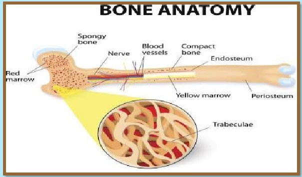
Osteons is the spindle-shaped unit present in the
compact bone. Osteocytes are present in the concentric layers of the matrix in
each osteon, called lamellae.There is a central marrow cavity made up of spongy
tissues (marrow).The yellow marrow contains fat, whereas red marrow produces
blood cells.
FUNCTIONS:Bones
have many functions. They support the body structurally,protect our vital
organs, and allow us to move. Also, they provide an environment for bone
marrow, where the blood cells are created, and they act as a storage area for
minerals, particularly calcium.
B) CARTILAGE:Cartilage
is mostly present in the embryonic stages and works as a
supporting skeleton. Most of the cartilage is replaced by bones in adults.

However, it supports some structures in adults too.
In humans,cartilage is present between the bones of the vertebral column, in
the
external ear, nose and hands. The cartilage consists
of chondrocytes cells,which are enclosed in a hard, rubbery matrix secreted by
them. They secretecollagen fibres also, which provide additional strength.
Functions:Cartilage is a flexible connective tissue
that keeps joint motion fluid by coating the surfaces of the bones in our
joints and by cushioning bones
against impact. It is not as rigid as bone, but is
stiffer and less flexible than
muscle tissue.Blood and lymph are fluid connective
tissues. Cells circulate in a liquid extracellular matrix. The formed elements
circulating in blood are all derived from hematopoietic stem cells located in
bone marrow.Erythrocytes (red blood cells), transport oxygen and some carbon
dioxide.
A) BLOOD:
Blood is a type of fluid connective tissue made up of various
cells present in the plasma. The blood contains red
blood cells (RBCs), white
blood cells (WBCs) and platelets.

FUNCTIONS :RBCs
have haemoglobin and transport oxygen.WBCs form a defence system and protect
from foreign antigens.
Platelets are important for blood clotting.
Plasma contains proteins, water, hormones, salts,
etc. to transport to different parts of the body.
B) Lymph:Lymph
drains into the blood and transports absorbed fat to the blood,
which cannot enter the bloodstream directly. Lymph
has white blood cells in
the liquid matrix. They help in getting rid of
toxins and waste materials. They
contain WBCs, which help in fighting infection.
LET US KNOW WHAT WE HAVE LEARNT!
A)MULTIPLE CHOICE QUESTIONS:
1. Under the
microscope, a tissue specimen shows cells located in spaces scattered in a
transparent background. This is probably
a) Loose connective tissue
b) bone
c) hyaline cartilage
d) tendon
2. Which
connective tissue specializes in storage of fat?
a) dense connective tissue
b) tendon
C) adipose tissue
d) reticular tissue
3. Ligaments
connect bones together and withstand a lot of stress.What type of connective
tissue should you expect ligaments to contain?
a) dense irregular connective tissue
b) areolar tissue
c) dense regular connective tissue
d) adipose tissue
4. In bone, the
main cells are
a) fibroblast
b) condroblast
c) lymphocytes
d) osteocytes
5. Connective
tissue is made of which three essential components?
a) Cells, ground substance, and protein fibers
b) Matrix, ground substance, and fluid
c) Cells, ground substance, and carbohydrate fibers
d) Collagen, ground substance, and protein fibers
B) Fill ups
1. Areolar tissue is a................... connective
tissue.
2. In comparasion to human erythrocytes, frogs
erythrocytes are.................
3. Tips of the nose and external ears are
...............
C) True /false
1. collagen is globular protein.
2.Ligament is modified white fibrous tissue.
3. Histamine is secreated by histiocytes.
A) MULTIPLE CHOICE QUESTION
Ans 1. c) hyaline cartilage
Ans 2. c) adipose tissue
Ans 3. c) dense regular connective tissue
Ans 4. d) osteocytes
Ans 5. a) Cells, ground substance, and protein
fibers
B) FILL UPS
1. loose
2. nucleated and with haemoglobin
3. cartilage
C) TRUE/FALSE
1. False (fibrous protein)
2. False (modified yellow elastic fibrous tissue)
3. False (mast cells)
1. Write a short note on Areolar tissue.
2. Differentiate between tendon and ligament.
3. Differentiate between bone and cartilage.
4. Discuss various functions of blood in animals.
1. Discuss in detail supportive connective tissue
its types and functions.
2. Discuss in detail fluid connective tissues, its
types and functions.
A75
INTRODUCTION:Dear
student, till now we have studied about Epithelial and Connective tissues and
their types. Today we will study about a Movement is one of the most important
characteristics of living organisms. Non-living objects do not move. The
movement of non-living organisms is induced while of living organisms are
autonomic (self-sustained).
BASIC TYPES OF MOVEMENTS:Amoeboid.
It is typical of Amoeba. It helps in food capture and change of place. This
type of movement is found in leucocytes.Ciliary Movement. |t is characteristic
way of ciliated protozoans such as Paramecium. Cilia of feeding apparatus of
Paramecium drive water and food. The cilia of Fallopian tubes and vasa
efferentia of human females and males, transport ova and spermatozoa,
respectively.Muscular Movement. This basic mechanism is used in the majority of
vertebrates, including humans. The universal
property of this mechanism is to exert a force by alternate contraction and
relaxation.Locomotion in Humans .Locomotion in Humans depends on the movement
of muscle fibres. Skeleton and joints
also help in locomotion.

MUSCLES:In
humans muscles constitute about 40-50 percent of the total body weight. These
muscles are broadly classified into three categories:
1. Skeletal or Striped or Voluntary muscles
2. Smooth or Unstriped or Involuntary muscles
3. Cardiac or heart muscles
SKELETAL MUSCLES.
These muscles are found in the limbs, body wall, tongue,pharynx and beginning
of oesophagus. These muscles are under the control of animals will. These
muscles are normally attached to the skeleton. Potassium is the most abundant
mineral element in muscles. Muscles store glycogen. They have oxygen carrying
pigment MYOGLOBIN. Muscles contain ATP, Creatinine,Phosphocreatinine, Urea etc.

SMOOTH MUSCLES:
These are found in posterior part of oesophagus, stomach.
intestine. lungs. urinary bladder, blood vessels.
iris of eyes, dermis of skin and
arrector pili muscle of hair. Smooth muscles are
never connected with skeleton.
Action of these muscles is controlled by autonomic
nervous system and hence they
are NOT under the control of the animal's will.

CARDIAC MUSCLES:
The cardiac muscles are found in the wall of the heart and
in the wall of large veins (e.g. pulmonary and
superior vena cava) where these veins
enter the heart. These show the characters of both
unstriped and striped muscle
fiber. Each fibre is a long and cylindrical has a
definite sarcolemma.

STRUCTURE OF SKELETAL MUSCLE:Each
organised skeletal muscle in our body is made of a number of muscle bundles or
fascicles held together by a common collagenous connective tissue layer called
fascia.Each muscle bundle contains a number of muscle fibers and each muscle
fibre is lined by the plasma membrane called ET enclosing
he Muscle fibre is a syncitium as the sarcoplasm
contains many nuclei.The endoplasmic reticulum, i.e., sarcoplasmic reticulum of
the muscle fibres is the store house of calcium ions.A large number of
parallelly arranged filaments are present in the sarcoplasm called as.Each
myofibril has alternate dark and light bands on it and it is due to the
distribution pattern of two important proteins: ACTIN and MYOSIN
The light bands contain actin and is called I-band
or isotropic band, whereas the dark band called ‘A’ or anisotropic band
contains myosin.Actin filaments are thinner as compared to the myosin
filaments, hence are commonly called thin and thick filaments respectively.In
the centre of each ‘|’ band is an elastic fiber called ‘Z’ line which bisects
it.The thin filaments are firmly attached to the ‘Z’ line and the thick
filaments in the ‘A’ band are also held together in the middle of this band by
a thin fibrous membrane called ‘M'’ line.The portion of the myofibril between
two successive ‘Z’ lines is considered as the functional unit of contraction
and is called a Sarcomere
In aresting state, the edges of thin filaments on
either side of the thick
filaments partially overlap the free ends of the
thick filaments leaving the
central part of the thick filaments, and the central
part of thick filament, not
overlapped by thin filaments is called the ‘H’ zone.


FUNCTIONS OF MUSCLE TISSUE
MOVEMENT: Our body's skeleton gives enough rigidity
to our body that skeletal muscles can yank and pull on it, resulting in body
movements such as walking, chewing, running, lifting, manipulating objects with
our hands, and picking our noses.
MAINTENANCE of posture:
Without much conscious control, our muscles generate a constant contractile
force that allows us to maintain an erect or seated position, or posture.
RESPIRATION:
Our muscular system automatically drives
movement of air
into and out of our body.HEAT GENERATION:
Contraction of muscle tissue generates heat, which is essential for maintenance
of temperature homeostasis. For instance, if our core body temperature falls,
we shiver to generate more heat.
COMMUNICATION: Muscle tissue
allows us to talk, gesture, write, and convey our emotional state by doing such
things as smiling or frowning.CONSTRICTION OF ORGANS AND BLOOD VESSELS:
Nutrients move
through our digestive tract, urine is passed out of
the body, and secretions are
propelled out of glands by contraction of smooth
muscle. Constriction or relaxation of blood vessels regulates blood pressure
and blood distribution throughout the body.
PUMPING BLOOD:
Blood moves through the blood vessels because our heart tirelessly receives
blood and delivers it to all body tissues and organs.
LET US KNOW WHAT WE HAVE LEARNT!
PART: A VERY SHORT ANSWER TYPE
QUESTIONS
A. MULTIPLE CHOICE QUESTIONS:
Q1 Functional
unit of striated muscle is:
(a)band
(b)Z-line
(c) Myofilament
(d) Sarcomere
Q2 Myoglobin
occurs in:
(a) skeletal muscle
(b) smooth muscle
(c) Cardiac muscle
(d) Both a and b
Q3 Which of the
following is multinucleated?
(a) Nonstriated muscle
(b) Striated muscle
(c) Renal tissue
(d) Nervous tissue
Q4 Largest Smooth
muscle is present in:
(a) Leg
(b)Thigh
(c)Uterus of pregnant women
(d) Urethra
Q5 Muscles which
are immune to fatigue are:
(a) Skeleton muscle
(b) unstriped muscle
(c) cardiac muscle
(d) jaw muscle
B. FILLIN THE BLANKS:
1. In muscle fiber Ca** is stored in ;
2. Two important protein are and .
3. The ions of ............play an important role in
muscle contraction.
C. TRUE/ FALSE:
1. Actin is present in thin filament.
2. H-zone of striated muscle fiber represents both
thick and thin filaments.
ANSWER KEY: PART-A
A. MULTIPLE CHOICE QUESTIONS:
1.d Functional unit of Striated muscle is sarcomere.
2.a Skeletal muscle fibers are rich in myoglobin
pigment.
3.b Striated muscles are syncitial
4.b Thigh have largest smooth muscle fibre.
5.c Cardiac muscles always work tirelessly
B. FILL IN THE BLANKS:
1. Sarcoplasmic Reticulum
2. Actin, Myosin
3. Calcium
C.TRUE/ FALSE:
1. TRUE
2. FALSE Z zone have thin and thick filaments.
PART: B SHORT ANSWER TYPE QUESTIONS
Q1. Distinguish between cardiac and striated
muscles?
Q2. How are thick and thin filaments arranged in a
muscle fiber?
Q3. Define role of myoglobin in the muscles.
PART: C LONG ANSWER TYPE QUESTIONS
Q1.Elaborate difference between Striated, Smooth and
Cardiac muscle
fibre.
Q2. Briefly discuss role of following in
muscles;Actin Myosin
A76
Dear students, as we have previously studied about
Epithelial, Connective and
Muscular tissues, in this assignment we will study
about Nervous Tissue its
Types and Functions.
NERVOUS TISSUE:Nervous
tissue is ectodermal in origin. It exerts the greatest control
over the body’s responsiveness to changing
conditions. Nerve cells are excitable by external and internal stimuli.
COMPOSITION OF NERVOUS TISSUE:
Nervous tissue is formed of four types of
cells:Neurons, Nerve cells Neuroglia
Ependymal Cells Neuro-secretory cells.
A. NEURONS:A
neuron is a nerve cell with all its branches. It is the structural
and functional unit of nervous system. It is the
communication unit of nervous system. It may be elongated over 100 cm. Neurons
can detect and receive and conduct the nerve impulses to various parts of the
body.A neuron is formed of two main parts:
Cyton, Nerve processes —Dendrites and Dendrons.
Cyton:
It is also called perikaryon or soma or cell body. It is of variable
shape. Its granular cytoplasm is called neuroplasm.
Dendrites and Dendrons: These are one or more small
sized tapering processes. Each is highly branched to form the terminal
arborization.These are afferent in nature and conduct the impulses towards the
cyton.

MAIN FUNCTIONS OF NEURONS:Neuron
is the basic unit of the brain Specialized cell designed to transmit
information to other
nerve cells, muscles or gland cells.
It helps transmit nerve signals, or impulses, down a
long axon.The main part of neuron is called cell body; it contains all
of the important parts of the cell that allow it to
function properly.
B. NEUROGLIA OR GLIA CELLS:These
are non-nervous cells which lie between the neurons of the CNS, ganglia and
between photoreceptors of retina of eye.These protect and support the neurons
and form more than one half the volume of nervous tissue.The Neuroglia cells
are of three types:Microglial cells. . Oligodendrocytes

FUNCTIONS:These
are capable of division and help in wear and tear of the CNS.These are insulate
the adjoining neurons and prevent the lateral transmission of impulses.
These provide nutrition to the neurons.
These acts as phagocytes and eat up the
microbes.These help in memory processes.
B.EPENDYMAL CELLS:These
are cuboidal and ciliated epithelial cells which line the
cavities of brain and spinal cord. These form an
epithelium called ependyma. Their cilia move the CSF.

FUNCTIONS:It plays an important
role in the production and regulation of CSF.The apical surfaces are covered in
a layer of cilia, which circulates CSF around the CNS.
The apical surfaces also covered with microvilli,
which absorbs CSF (Cerebro Spinal Fluid)
C. NEUROSECRETORY CELLS:These
are special type of neurons of the hypothalamus of brain. These are endocrine
in function and secret neurohormones which are carried
by the blood. Ex. TSH, STH, FSH, etc.

FUNCTIONS:It is also a
type of neuron or nerve cell, whose function is to translate
neural signals into chemical stimuli.

(A). Multiple choice questions:
(i). The
structural and functional unit of nervous system is....
(a). Cell
(b). Tissue
(c). Neuron
(d). Nephron
(ii). Which cells
lie between the neurons of the CNS, ganglia and between photoreceptors of
Retina of eye
(a). Neurosecretory cells.
(b). Ependymal cells
(c). Neuroglia cells.
(d). Cyton
(iii). Which
cells plays an important role in the production and regulation of CSF.
(a). Ependymal cells
(b). Glia Cells
(c). Neurosecretory cells
(d). Dendron
(iv). Neuron is
the basic unit of
(a). Heart
(b). Brain
(c). Kidneys
(d). Lungs
(v). Cyton is
also called.
(a). Perikaryon
(b). Nephron
(c). Dendron
(d). None of above
(B). True or False:
(i). Neuron is the structural and unctional unit of
nervous system.
(ii). The main function of neurosecretory cells is
to translate neural
signals into chemical stimuli.
(C). Fill in the blanks:
(i). cells are special type of neurons of the
hypothalamus.
of brain.
(ii). CSF stands for .
(iii). cells are endocrine in function.
ANSWER KEY: PART-A
(A). Multiple choice questions:
(i). (c) Neuron Neuron is the structural and
functional unit of nervous
system.
(ii). (c). Neuroglia cells. Cells lie between the neurons
of the CNS,
Ganglia and between Photoreceptors of retina of eye.
(iii) (a). Ependymal cells. plays an important role
in the production and
regulation of CSF.
(iv) (b). Brain. Neuron is the basic unit of brain,
structural and
functional unit of nervous system.
(v) (a).Perikaryon. It is also called perikaryon or
soma or cell body.
(B). True or False:
(i). True. Neuron is the structural and functional
unit of nervous system.
(ii). True. It translate neural signals into
chemical stimuli.
(C). Fill in the blanks:
(i). Neurosecretory cells.
(ii). Cerebro Spinal Fluid
(iii). Neurosecretory
(i). Define the term neuron. Write its types.
(ii). Draw a structure of typical neuron.
(iii). Write main functions of (i). Neurons (ii).
Neurosecretory cells.
(iv). Write short note on. Cyton and Dendrites,
Dendrons.
(i). Briefly explain types of neurons with its main
functions.
A77
COCKROACHES
are the most wide spread of all insects being worldwide in distribution.
Cockroaches are brown or black bodied animals that are included in class
Insecta of Phylum Arthropoda.
Phylum: Arthropoda
Class: Insecta
Subclass: Pterygota
Order: Dictyoptera
Family: Blattidae
Genus: Periplaneta
Species: americana
MORPHOLOGY Colour:
Brownor Black. (Bright yellow, red and green coloured ones
have also been reported in tropical regions)Size:
Ranges from % inches to 3 inches (0.6-7.6 cm.
Grossly: They have long antenna, legs and flat extension of the upper body wall that conceals head.Behaviour: Nocturnal omnivores live in damp places throughout the world. They have become residents of human homes and thus is serious pests and vectors of several diseases.

The adults of the common species of cockroach, INE
GE are about 34-53 mm long with the wings that
extend beyond the tip of the abdomen in males.The body is segmented and
divisible into three distinct regions
HEAD, THORAX and ABDOMEN.The entire body is covered by a hard chitinous exoskeleton (brown in colour). In each segment, exoskeleton has hardened plates called SCLERITES (Tergites dorsally and Sternites ventrally) that are joined to each other by a thin and flexible articular membrane (Arthrodial Membrane).

Triangular in shape and lies
anteriorly at right angles to the
longitudinal body axis.Formed by fusion of six
segments and shows great mobility in all directions due to flexible neck.
A pair of compound eyes and a pair of thread like
antennae which have sensory receptors that help in monitoring the
environment.Biting and chewing type of mouth parts.
Mouthparts-
a) Labrum(upper lip),
b) a pair of mandibles,
c) a pair of maxillae,
d) Hypopharynx (acting as tongue).

It consists of three parts: Prothorax, Mesothorax,
Metathorax.Connected to the head by short extension of the prothorax
known as the neck.Each thoracic segment bears.a pair
of walking legs.First pair of wings ( forewings) arises from mesothorax and
second pair(hindwings) from metathorax.
Forewings (called teq miria) are opaque dark and
leathery and cover the hind wings when at rest The hind wings are transparent,
membranous and are used in
flight.

Females:
The 7" sternum is boat shaped and together with 8" and 9 sterna forms
a brood or genital pouch whose anterior part contains female gonopore,
spermathecal pores and collateral glands.
Males:
The genital pouch lies at the hind end of the abdomen bound dorsally by 9t* and
10* terga and ventrally by the 9%
sternum. It contains dorsal anus, ventral male genital pore and gonapophysis.Males bear a pair of short thread like [J which are absent in females.
In both sexes, the 10% segment bears a pair of joined filamentous structures called

COCKROACH is RR. However true coelom occurs only in
embryonic stage. The body cavity of adults is filled
with J GR and is called a
The alimentary canal is divided into three regions:The mouth opens into a short tubular EE leading to a narrow tubular passage called IEE which inturn opens into a sac like structure called J used for storing of food.

The crop is followed by gizzard or proventriculus, which
has six
highly chitinous plates called teeth. It helps in
grinding the food particles.
Atjunction of foregut and midgut, a ring of 6-8
blind tubules called hepatic or gastric caecae is present, which secretes
digestive
juice.At junction of midgut and hindgut is present another ring of 100-150 yellow coloured thin filamentous Malpighian tubules, which removes excretory products from hemolymph.
The hindgut is divided into
ileum, colon and rectum. The rectum
opens out through anus.
Blood vascular is an open type. Blood vessels are poorly developed and open into haemocoel.

Visceral organs are located in haemccoel and bathed
in haemolymph, which is composed of colourless plasma and haemocytes.
Heart consists of elongated muscular tube lying along mid dorsal line of thorax and abdomen. It is differentiated into funnel shaped chambers with ostia on either side. Blood from sinuses enter heart through ostia and is pumped anteriorly to sinuses again.It consists of a network of trachea, that opens through 10 pairs of small holes called spiracles present on the lateral side of the body.Tracheal tubes are divided into tracheoles and carry oxygen from air to all the parts.The opening of the spiracles is regulated by the sphincters.

Cockroach is jificetelic.Excretion is performed by
Malpighian tubules. Each tubule is lined by glandular and ciliated cells. They
absorb nitrogenous waste products and convert them into uric acid which is
excreted out through the hindgut.

It consists of a series of fused, segmentally
arranged ganglia joined by paired longitudinal connectives on the ventral
side.Three ganglia lie in the thorax, and six in the abdomen.The head holds a
bit of a nervous system while the rest is situated along the ventral side of
the body.Brain is represented by supra-oesophageal ganglion which supplies
nerves to the antennae and eyes.Sense organs- ne
Each eye consists of about 2000 hexagonal ommatidia. With help of several ommatidia, a cockroach receives several images of an object. This kind of vision is known as i with more sensitivity and less resolution.

Cockroaches are dioecious and both sexes
have well developed reproductive organs.Male consists of a pair of testes one
lying on each lateral side in the 4%"-6t* abdominal segments. From each
testis arises a thin vas deferens, which opens into ejaculatory duct through
seminal
vesicle. The ejaculatory duct opens into male
gonopore situated ventral to anus.
Accessory reproductive organ- Mushroom shaped gland present in the 6%-7" abdominal segments.External genitalia- Male gonapophysis or phallomere.Sperms are stored in the seminal vesicles and are glued together in form of bundles called as spermatophores which are discharged during copulation.

Females consists of two large
ovaries, lying laterally in 2"¢ -6"
abdominal segments. Each ovary is formed of a group of eight ovarian tubules or ovarioles, containing a chain of developing ova.Oviducts of each ovary unite into a single median oviduct(also called vagina) which opens into the genital chamber. A pair of spermathecal is present in the 6 segment which opens into the genital chamber.

Sperms are transferred
through spermatophores.Their fertilised eggs are encased in capsules called
Bjaaaaa
(Dark reddish to blackish brown capsule, about 3/8”
(8mm)long), which are dropped or glued to a suitable surface.On an average,
females produce 9-10 oothecae, each containing: 14-16 eggs.Development is
paurometabolous, meaning there is development through nymphial stage. The nymph
looks very much like adults.
The nymph grows by moulting 13 times to reach the
adult form.The next to last nymphal stage has wing pads but only adult
cockroaches have wings.
A.) MULTIPLE CHOICE QUESTIONS:
1.) The body of
cockroach is divided into how many parts?
a) Two
b) Three
c) One
d) Undivided
2.)Which of the
following is not a mouthpart of the cockroach?
a.) Labrum
b.) Hypopharynx
c.) Sclerites
d.) Maxillae
3.) Which of the
following segment in female’s abdomen is boat
shapes and takes
part in brood formation?
a.) 6
b.) 7
c.) gm
d.) 10%
4.) Hepatic
caecae are present at junction of:
a.) Hypopharynx and foregut
b.) Foregut and midgut
c.) Midgut and hindgut
d.) Foregut and hindgut
5.) Ovaries are
present in which of the following segments?
a.) 1% -7'
b.)2nd.6"
c.) gth_gth
d.)5t-gtn
B.) FILL UPS:
1.)In both sexes, the 10 segment bears a pair of
joined filamentous
structures called
2.). Heart is differentiated into funnel shaped
chambers with
on either side.
3.) Brain is represented by which supplies nerves to
the
antennae and eyes.
C.) TRUE OR FALSE:
1.) The hindwings are dark, leathery and opaque.
2.) Accessory reproductive organ in males (Mushroom
shaped
gland) is present in the 6-7 abdominal segments.
ANSWER KEY: PART (A)
A) MULTIPLE CHOICE QUESTIONS :
1. b) Three parts
2 c)Sclerites
3. b) 7 segment
4 b) Foregut and midgut
5. b) 2nd.6th
B) FILL UPS:
1. Anal cerci
2. Ostia
3. Supra-oesophageal ganglion
C) TRUE/ FALSE:
1. False
2. True
PARTB: SHORT ANSWER TYPE QUESTIONS
1.) Name the mouthparts of the cockroach.
2.) Write a short note on the Gastrointestinal
system of the cockroach.
3.) Write salient features of the thorax of
cockroach.
PARTC: LONG ANSWER TYPE QUESTIONS
1.) What are the major differences between male and
female reproductive
system of a cockroach?
2.) Draw a well labelled diagram of female
reproductive system of
cockroach.
A78
RECAPITULATION:Dear
students in the chapter “STRUCTURAL ORGANISATION IN ANIMALS.
you have read all the topics in detail. As this
chapter deals with the internal
structure of animals, we have learnt that animals
have cells as the basic unit,
cells are organized into tissues and in tum the
tissues are organized into organsand organs are organized to form organ systems
and then into complete organism.
Now let us do NCERT book questions;
Q2. Answer the
following.
(i) What is the function of nephridia?
Solution: (i) Nephridia are excretory organs of
earthworm, which perform the
function of excretion and osmoregulation. Nephridia
regulate the volume and
composition of the body fluids.

Q6. What are the
ceilular components of blood?
Solution: Blood is a fluid connective tissue. It is
composed of EERE Ge ee Cellular components of blood (blood corpuscles) OR See
constitute about 45% of blood volume.Three types of blood cells are:Ree. they
are most abundant blood cells.Normal RBC count is 5-5.5 million/mm? in males
and 4.5-5 million/mm? in
females) RBCs help in transport of gases and
maintain blood pH. The normal WBC count is 5000- 6000/mmé of blood. They are
involved in immune response of body and act as soldiers and scavangers Ee.
There are about 2,50,000 platelets/mm® of blood. They are involved in blood
clotting.
Q7. What are the
following and where do you find them in animal body?
(a) Chondrocytes
(b) Axons.
(c) Ciliated epithelium
Solution: (a) Chondrocytes —
Chondrocytes are the only cells found in cartilage. They are present in spaces
called lacunae and they produce andmaintain the matrix of cartilage. Bending
ability of cartilage is due to chondrocytes. Cartilage is present at tip of
nose, pinna of ear, epiglottis etc.
(b) Axon
— Axon is one of the processes of neuron, which is the structural and
functional unit of nervous system. It conducts impulses away from the

cyton. Neurons (nerve cells) are present in brain and
spinal cord.
(c) Ciliated epithelium
— If the columnar or cuboidal cells bear cilia on their
free surface they are called ciliated
epithelium.Their function is to move particles or mucus in a specific direction
over the epithelium.They are mainly present in the inner surface of hollow
organs like bronchioles and Fallopian tube.
Q 8. Describe
various types of epithelial tissues with the help of labelled
diagrams.
Solution: Epithelial tissue is a tissue made of one
or more layers of compactly arranged cells that covers external surface and
internal free surface of body organs and which is underlined by a basement
membrane.The various types of epithelial tissue along with the diagram are given
below:

(i) Siviple epithelium
; It is composed of single layer of cells resting on
basement membrane.Simple epithelium generally occurs
over secretory and absorptive surfaces.imple epithelium is of several types.
(a) Squamous epithelium:
It consists of single layer of flat cells, tightly linked
together and have centrally located oval or
spherical nucleus.It is also called pavement epith lium. It is found in walls
of "blood vessels, air sacs of lungs, and lining of eye lens.

(b) Cuboidal epithelium:
Cells of cuboidal epithelium are as tall as wide, with centrally placed
nucleus.Its main functions are secretion and absorption. It lines sweat gland,
thyroid follicles, salivary glands.Brush bordered cuboidal epithelium, i.e.,
cells having microvilli on their free surface lines proximal part of
uriniferous tubule,pancreatic duct, testis and ovary.

(c) Columnar epithelium:
Cells are with basaily located nucleus.It helps in secretion and absorption.It
occurs in lining of intestine, stomach, gall bladder.

(d) Ciliated epithelium:
Free surface of columnar and cuboidal cells are
covered with cilia.Cilia help in moving fluids,
particles, mucus, etc. in a specific direction.It occurs in the inner surface
of Fallopian tubules, nasal passage,bronchioles.

(e) Pseudostratified epithelium:
It consists of single layer of cells but some
cells are shorter than others.Due to difference in
size of cells, the epithelium appears 2-3 layered.Pseudostratified columnar
epithelium occurs in urethra and parotid salivary gland. Pseudostratified
columnar ciliated epithelium (only larger
cells ciliated) occurs in lining layer of nasal’
chambers, trachea and large bronchi.
It helps in moving mucus and foreign particles.

(ii) Compound Epithelium / Stratified
Epithelium:It is multilayered epithelium where cells of only
the lowermost or basal layer are in contact with basement membrane.It provides
protection against mechanical and chemical stresses and has
limited role in secretion and absorption.
It covers dry surface of skin, moist surface of
buccal cavity, pharynx,etc.
Different types of compound epithelium are:
[he cells of outer layer are flattened
and squamous while the inner layers are cuboidal
cells.Itis of two types: Non- kKeratinised lining oesophagus, pharynx, buccal
cavity, cornea, vagina and anal canal and keratinised (comified):
forming epidermis of skin, hair, horn and nail.

he
outer layer of cuboidal cells and
basal layer of columnar cells. It lines ducts of
sweat glands, large salivary and
pancreatic ducts. Both upper and basal layers are
made of columnar cells, e.g., epiglottis covering, part of urethra.
Pe. Outer layer consists of ciliated
columnar cells and basal layer of columnar cells, e.g., larynx.This is stratified epithelium which contains cuboidal or columnar shaped cells, which are thin and stretchable.No basement membrane is present.It lines the inner surface of renal calyces, urinary bladder, ureter.

It consists
of specialised epithelial cells which synthesise intracellular macromolecules
(protein in pancreas, lipids in adrenal glands, glycoprotein in salivary glands
and all the three in mammary glands)


Q10. Mark the odd
one in each series.
(a) Areolar tissue; blood; neuron; tendon
(b) RBC; WBC; platelets; cartilage
(c) Exocrine; endocrine; salivary gland; ligament
(d) Maxilla; mandible; labrum; antennae
(e) Protonema; mesothorax; metathorax; coxa.
Solution:(a) Neuron: Areolar
tissue, blood and tendon are connective tissues while
neuron is a part a nervous tissue.
(b) Cartilage:
RBC, WBC and platelets are parts of vascular connective tissue
while cartilage is skeletal connective tissue.
(c) Ligament:
Ligament is a connective tissue.
(d) Antennae:
Maxilla, mandible and labrum are mouth parts of cockroach
while antennae are sense organs.
(e) Protonema:
Protonema is a filamentous juvenile stage in life cycle of
Bryophytes, while mesothorax, metathorax and coxa
are appendages of cockroach.

Q14. (b) Mention
the function of the MALPIGHIAN TUBULES.
Solution. MALPIGHIAN TUBULES are the excretory
organs in Cockroach.
These lie openly in the haemolymph. They extract
excretory materials from
the haemolymph and open up at the junction of mid
gut and the hind gut in the
food canal. They are around 150 in number.
A79
Dear students, as we have already studied about
various tissues found in animals now we will sum up the chapter with important
diagrams from the chapter and run through the chapter with the help of a test.
The structure of the cells varies according to their
function. Therefore, the
tissues are different and broadly classified into
four types:
1. EPITHELIAL TISSUE
2. CONNECTIVE TISSUE
3. MUSCULAR TISSUE
4. NEURAL TISSUE
EPITHELIAL TISSUE
An epithelium is a tissue composed of one or more layers of cells covering the
external and internal surfaces of various body parts. Epithelial tissue also
forms glands.There are two types of epithelial tissues namely:
1. SIMPLE EPITHELIUM
2. COMPOUND EPITHELIUM
These tissues show different types on the basis of
cell shape and number of layers they form. For eg. Squamous epithelium,
Cuboidal epithelium,Columnar epithelium, Ciliated epithelium, Cuboidal
epithelium and Glandular epithelium.

CONNECTIVE TISSUE
Connective tissues, as the name implies, support and connect different tissues
and organs of the body. Connective tissue is made up of a few
cells present in the intercellular framework of
protein fibres secreted by
the cells, known as a es,
TYPES OF CONNECTIVE TISSUE:
1. LOOSE CONNECTIVE TISSUE
2. SPECIALISED CONNECTIVE TISSUE
3. DENSE CONNECTIVE TISSUE

MUSCULAR TISSUE In humans muscles constitute about
40-50 percent of the total body weight.These muscles are broadly classified
into three categories:
1. SKELETAL OR STRIPED OR VOLUNTARY MUSCLES
2. SMOOTH OR UNSTRIPED OR INVOLUNTARY MUSCLES
3. CARDIAC MUSCLES

NEURAL TISSUE
Nervous tissue is ectodermal in origin. It exerts the greatest control over the
body’s responsiveness to changing conditions. Nerve cells are excitable by
external and internal stimuli.Nervous tissue is formed of: Neurons and Nerve cells, Neuroglia

Cockroaches are the most wide
spread of all insects being worldwide in distribution. Cockroaches are brown or
black bodied animals that are
included in class Insecta of Phylum Arthropoda.
Phylum: Arthropoda
Class: Insecta
Subclass: Pterygota
Order: Dictyoptera
Family: Blattidae
Genus: Periplaneta
Species: americana


Cockroach is coelomate. However true coelom occurs
only in embryonic stage. The body cavity of adults is filled with haemolymph
(blood) and is called haemocoel. Internal anatomy
shows following vital
organ systems


A.) MULTIPLE CHOICE QUESTIONS:
1. Gap, tight and
adhering junctions are found in:
a) Epithelial tissue
b) Connective tissue
c) Muscular tissue
d) Neural tissue
2. Ligaments
connect bones together and withstand a lot of stress.
What type of connective tissue should you expect
ligaments to contain?
a) Dense irregular connective tissue
b) Areolar tissue
c) Dense regular connective tissue
d) Adipose tissue
3. Muscles which
are immune to fatigue are:
a) Skeleton muscle
b) Unstriped muscle
c) Cardiac muscle
d) Jaw muscle
4. The structural
and functional unit of nervous system is:
a) Cell
b) Tissue
c) Neuron
d) Nephron
5. The body of
cockroach is divided into how many parts?
a) Two
b) Three
c) One
d) Undivided
B)FILL UPS:
1. Areolar tissue is a connective tissue.
2. In comparison to human erythrocytes, frog’s
erythrocytes are .
3. Brain is represented by which supplies nerves to
the antennae and eyes.
C) TRUE /FALSE:
1. Actin is present in thin filament.
2. Accessory reproductive organ in males (Mushroom
shaped gland) is present in the 6-7" abdominal segments.
ANSWER KEY - PARTA
A)MULTIPLE CHOICE QUESTIONS:
1. a) These are junctions which help in joining of
adjoining cells.
2. c) Dense regular connective tissue.
3. c) Cardiac muscles work tirelessly.
4. c) Neuron- helps in transmission nerve impulses.
5. b) Cockroach is divided into three parts.
B)FILL UPS:
1. Loose
2. Nucleated with haemoglobin
3. Supra-oesophageal ganglion
C) TRUE OR FALSE:
1. TRUE
2. TRUE
PART B: SHORT ANSWER TYPE QUESTIONS
1. Name the mouthparts of the cockroach.
2. How are thick and thin filaments arranged in a
muscle fiber?
3. Write a short note on specialised connective
tissue.
PART C: LONG ANSWER TYPE QUESTIONS
1. Draw a well labelled diagram of female
reproductive system of cockroach.
2. Elaborate difference between Striated, Smooth and
Cardiac muscle fiber.
3. Describe various types of epithelial tissues with
the help of labelled
diagrams.
