6 ANATOMY OF FLOWERING PLANTS
CHAPTER NO.6 ANATONY OF FLOWERING PLANTS
A65
INTRODUCTION:TISSUES:
Cells of plants exhibit great variation in size and structure.A group of cells
performing essentially the same function and commonly of similar structure is
called a tissue.
Depending upon the plant organ, various tissues are
distributed in
characteristics patterns within the plant body.Plant
Tissues are constituted of three basic types of systems.
1)Epidermal 2) Ground 3) Vascular
Let us discuss today Epidermal Tissue and Stomata:
EPIDERMIS: It is the outermost layer of all organs of primary plant body.sually,It is made up of single layer of cells. However in some plants it is multi-layered,Ex.Nerium.
The Epidermis of differs in origin, structure and function and called as epiblema layer.
It is made up of elongated, compactly arranged cells.Cells constituting epidermal cells are parenchymatous cells.
With little
quantity of cytoplasm lining the cell wall with a large vacuole.The outer wall
of epidermal cells contain a fatty substance called cutin and form cuticl on
the epidermis. On the surface of cuticle, a waxy coating may also be present.
STOMATA:
These
are the structures present in the epidermis of leaves.
Stomata help in gaseous exchange at the time of
respiration and
photosynthesis.They are minute pores found on the epidermis of leaves, stem etc.Stomata transpiration takes place through stomata. They are two .dney shaped guard cells which bound a minute elliptical pore in a stoma.
The guard cells are modified epidermal cells. The wall of guard cells near the pore is thick. The outer wall is thin, elastic and semipermeable.In monocots the guard cells are Dumb-bell in outline.The guard cells are filled with chloroplasts.Both kidneys shaped and dumb-bell shaped guard cells have been reported in Cyperus.Average length of stomata is 20 micro meter to 28 micro meter and breadth 5 micrometer.
The opening and closing of stomata is controlled by guard cells.When water flows into the guard cells, they swell up and the curved surfaces cause the stomata to open.When the guard cells lose water, they shrink and become flacid and straight closing the stomata.
Part: AVERY SHORT ANSWER TYPE
QUESTIONS:
(A).Multiple choice questions:
(i). The study of
tissue is called...
(a). Biology
(b).Histology
(c).Microbiology
(d).Zoology
(ii).Group of
cells which performs specific functions is called.....
(a). Tissue
(b).Neuron
(c).Cytoplasm
(d).Nephron
(iii). The tiny
pores present on the epidermis layer of leaves which helps in exchange of gaseous
called
(a).Lenticels
(b).Stomata
(c).Guard cells
(d).Nucleus
(iv). The outer
wall of epidermal cells contains a fatty substance called....
(a). Cutin
(b).Melanin
(c). Cuticle
(d).None of above
(v).Epiblema of
roots is equivalent to..
(a).Pericycle
(b).Endodermis
(c).Epidermis
(d).Stele
(B). State True or False:
(i). Study of tissue is called zoology.
(ii). Stomata help in gaseous exchange at the time
of respiration and photosynthesis.
(C).Fill in the blanks.
(i). The outer wall of epidermal cells contain a
fatty substance called.........
(ii). The opening and closing of stomata is
controlled by..........
PE
A. Multiple choice questions:
(i). (b) histology: study f tissue is called
histology.
(ii). (a) tissue: group of cells is called tissue.
(iii). (b) stomata: tiny pores present on the
epidermis layer of leaves.
(iv). (a) cutin: outer wall of epidermal cell,
contains fatty substance
B.True or False.
(i). false: Study of tissue is called histology
(ii). True: Stomata help in exchange of gaseous.
C.Fill Up:
(i).Cutin
(ii).Guard cells
(B).Short Answer Type Questions:
1. Define the terms:
(a). Tissue.
(b).Stomata
2. Write short note on cause of opening and closing
of stomata.
3. Plant Tissues are constituted of three basic
types of systems, name them.Explain Epidermis.
4. Give the functions of guard cells, in plants.
(c).Long Answer Type Questions.
1. Draw a well labeled diagram of stomata. Which
shows opening and _ closing of stomata. Also write the main functions of
stomata.
A66
INTRODUCTION:Dear students
you have read about different types of plant tissues. Lets now consider how
tissues vary depending on their location in the plant body. Their structure and
function would also be dependent on location. Today we will discuss about
ground tissue system and vascular tissue system.
GROUND TISSUE SYSTEM:
All tissues except epidermis and vascular bundles constitute Ground tissue.This
system contains three cell types called parenchyma,collenchyma and
sclerenchyma.
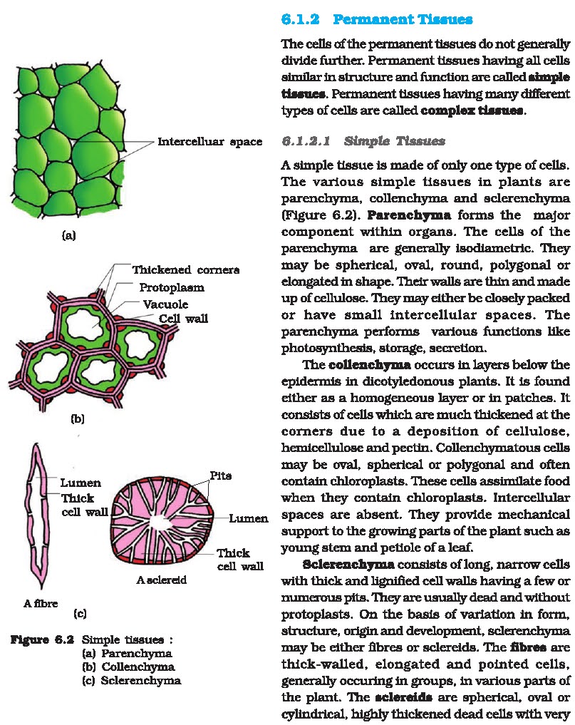
PARENCHYMA: Cells are found
in all tissue types.They are living cells ,generally capable of further
divisions, and have a thin primary cell wall. These cells have a variety of
functions.The apical and lateral meristematic cells of shoots and roots provide
the new cells required for growth.Food production and storage occurs in the
photosynthetic cells of the leaf and stem called mesophyll cells, storage
parenchyma cells form the bulk of most fruits and vegetables.
COLLENCHYMA:
These are living cells similar to parenchyma cells, except that they have much
thicker cell walls and are usually elongated and packed into long rope like
fibres. They are capable of stretching and provide mechanical support in the
ground tissue system of the elongating regions of the plant. Collenchyma cells
are especially common in sub epidermal region of stem. Based on cell thickness
and arrangement these are of four types; angular, annular, lamellar, and
lacunar.
SCLERENCHYMA:Like
collenchyma it has strengthening and supporting functions. However they are
usually dead cells with thick lignified secondary cell walls that prevent them
from stretching as the plant grows. Two common types are fibres,which often
form long bundles and sclereids, which are shorter branched cells found in seed
coat and fruit. Based on functions and morphology these are classified as
under;
VASCULAR TISSUE SYSTEM: The phloem and the xylem together form a continuous vascular system throughout the plant In young plants they are usually associated with a variety of other cell types in vascular bundles. Both phloem and xylem are complex tissue. Their conducting elements are associated with parenchyma cells that maintain the elements and exchange materials with them. In addition, groups of collenchyma and sclerenchyma cells provide mechanical support.
In dicotyledonous stems
cambium is present between phloem and xylem. Such vascular bundles are called
open vascular bundles.In monocotyledons the vascular bundles have no
cambium,hence these are called closed vascular bundles.
PHLOEM:
Phloem is involved in the transport of organic salutes in the plant. The main
conducting cells are aligned to form tubes called sieve tubes. The sieve tube
elements at maturity are living cells, nterconnected perforations in their end
walls formed from enlarged and modified plasmodesmata called sieve plates.These
cells retain their plasma membrane, they have lost their nuclei and much of
their cytoplasm.They therefore rely on associated companion cells for their
maintenance. These companion cells have the additional function of actively
transporting soluble food molecules into and out of sieve tube elements through
porous sieve areas in the wall
XYLEM:
Xylem carries water and dissolved ions in the plant. The main conducting cells
are the vessel elements ,which are dead cells at maturity that lack a plasma
membrane The cell wall has been secondarily thickened and heavily lignified.
The vessel elements are closely associated with xylem parenchyma cells. which
actively
transport selected solutes into and out of the
elements across the parenchyma cell plasma membrane.the structural elements of
xylem are tracheids, trachea, xylem fibre, xylem parenchyma and rays.
VASCULAR BUNDLES:
Roots usually have a single vascular bundle, but dicot stems
have several bundles. These are arranged with strict
radial symmetry in dicots but they are more irregularly dispersed in monocots.
Let us know what we have learnt
PART-A [VERY SHORT ANSWER TYPE
QUESTIONS]
Multiple choice questions:
1. Food
production and storage occurs in the photosynthetic cells of the leaf
and stem which are called as:
a) Xylem fibres
b) Mesophyll cells
c) Sieve plates
d) Both A and B
2. Angular type
of cells are found in:
a) Xylem
b) Phloem
c) Collenchyma
d) Parenchyma
3. Vascular
tissue system consists of:
a) Xylem
b) Phloem
c) Sclerenchyma
d) BothA andB
4. Plasmodesmata
are found in:
a) Phloem
b) Xylem
c) BothA and B
d) None of these
5. Vascular
bundles with strict radial symmetry are found in:
a) Dicot stem
b) Monocot stem
c) Parenchyma
d) None of these
Fill in the blanks:
1. All tissues except epidermis and vascular bundles
constitute.........tissue
system.
2. Angular, annular, lamellar, and lacunar are types
of................26
3. Tracheids are present in..................
True/False
1. Xylem carries water and dissolved ions in the
plant.
2. Parenchyma, collenchyma and sclerenchyma form
vascular tissue system.
3. Xylem parenchyma cells actively transport
selected solutes into and out of
the elements across the parenchyma cell of plasma
membrane.
ANSWER KEY: [PART-A]
A) Multiple choice questions:
1. a(Mesophyll cell are found in ground
tissue-parenchyma)
2. c(collenchyma consists of four types of cells
,angular form is one of them)
3. d(Sclerenchyma is a ground tissue)
4. a(Sieve plates in phloem are also called
plasmodesmata)
5. a(ln monocots these are irregularly dispersed)
B) Fill in the blanks:
1. Ground tissue
2. Collenchyma
3. Xylem
C) True/False:
1. True
2. False (Xylem and phloem form vascular tissue
system)
3. True
PART-B [SHORT ANSWER TYPE QUESTIONS]
1. Write the types of sclerenchyma based on its
function and morphology.
2. What are the structural elements of xylem?
3. What is the function of sclerenchyma cells?
PART-C [LONG ANSWER TYPE QUESTIONS]
1. Give an account of vascular tissue system in
plants.
2. Write a note on ground tissue system in plants
A67
INTRODUCTION:Study
of internal structure of the plants is known as ANATOMY.N.Grew, the father of
plant anatomy was first to use term tissue.A tissue is group of cells having common,
same method of development
and similar structure and function. The cells in a
tissue may be all of one similar
type or of more than one dissimilar types. A tissue
is formed in response to a
basic division of labour. Anatomy deais with
internal organization of plants.
In plants Anatomy and Histology have same
meaning.Tissues are classified into two types on the basis of stage of
development:
MERISTEMATIC TISSUE
PERMANENT TISSUE We have already discussed in detail
about different types of tissues in previous Daily Dose Assignments. Now let us
discuss in detail about Anatomy of Monocot and Dicot Root.ANATOMY OF MONOCOT
ROOT In transverse section, a monocot root (e.g Wheat, Maize) shows three
zones:
1. Epidermal zone.
It consists of epiblema. Cuticle is absent. It consists of single layer of thin
walled cells. There are no intercellular spaces. Some of the
epiblema cells have tubular outgrowths called Root
Hairs. The cells producing
root hair are called Trichoblasts. The epiblema is
protective as well as bsorptive
in function.
2. Cortical zone.
It is wider in monocot roots. It consists of parenchymatous
cortex. In older roots when epidermis disintegrates,
outer layers of cortex
become thick walled and suberised to form exodermis
for protection and
prevention of leakage of water. The innermost layer
of cortex is called
endodermis made up of barrel shaped cells without
intercellular spaces.
3. Stellar zone.
It consists of single or multilayered pericycle from which
only lateral roots arise. Below it is 8 or more
(polyarch condition)alternately and radially arranged xylem and phloem bundles
and Parenchymatous Pith in the center.
ANATOMY OF DICOT ROOT
In Transverse Section of Dicot Root (e.g. Gram, Sunflower, Tomato, Pea)shows
three zones of tissues:
1. Epidermal zone
, the outermost zone,consists of Epiblema (piliferous layer)or rhizodermis:
Epidermis without cuticle and stomata, bearing unicellular root hairs and
taking part in absorption of water is called epiblema. The cells are thin
walled andtubular. The cells producing root hair are called Trichoblasts. Root
hairs are actually thin walled out growths of
epidermal cells. This epiblema is
protective as well as absorptive in function.
2. Cortical zone.
It consisting of thin walled parenchymatous, round oval or
angular cells forming cortex. Barrel shaped cells
without intercellular spaces form Endodermis, the specialised innermost layer
of cortex. Small but abundant intercellular spaces are present in cortex.
Cortex is never sclerenchymatous in root. Endodermis bears Casparian strips on
anticlinal walls on its cells. The cells of endodermis opposite to protoxylem
are thin walled and without lignin, called passage or transfusion cells.
3. Stellar zone.
Parenchymatous single layered Pericycle is present below
endodermis. It forms the outer boundary of stele or
vascular strand. It forms
lateral roots, part of vascular cambium above xylem
region and cork cambium by
Dedifferentiation. Vascular strand consists of 2-6
radial bundles each of xylem and phloem. Phloem bundles are in the form of
small oval patches in contact with the pericycle. Xylem is Exarch as protoxylem
is on the outer side in contact with the pericycle. Xylem bundles are four in
Sun flower root, so Tetra arch. In between adjacent Xylem and Phloem, the
conjunctive parenchyma is present.The different xylem bundles may come in
contact centrally to form Metaxylem plate, and pith may be absent.
Let us know what we have learnt!
a) Multiple Choice questions:
1. In plants,
anatomy is also called:
(a) Dendrochronology
(b) Histology
(c) Cytology
(d) internal cytology
2. Root Cap in
dicots is formed from:
(a) Ground meristem
(b) Procambium
(c) Protoderm
(d) Calyptrogen
3. Pericycle of
Dicot root does not take part in formation of:
(a) Cambium
(b) Lateral roots
(c) Root Hair
(d) Cork Cambium
4. Casparian
strip occurs in the roots on radial walls of:
(a) Exodermis
(b) Passage cells
(c) Endodermis
(d) Pericycle
5. Xylem vessel
shows :
(a) Two way direction of flow of water
(b) One way direction of flow of water
(c) Both of these
(d) None of these.
b) Fill in the blanks:
1. is regarded as father of Plant Anatomy.
2. Xylem vessels are generally .
3. Cortex is very in monocot root.
c) True/False:
1. Root Cap is cushion of parenchymatous tissue for
protection.
2. The root hairs are not derivatives of epidermal
cells.
ANSWER KEY: PART-A
A) Multiple choice questions
1. b (Histology is the other name for anatomy)
2. c(In dicots the root cap is derived from the
protoderm. )
3. c Pericycle of dicot root doesn't involves in
formation of root hairs.
4. c Radial wall of Endodermis bears Casparian
strips.
5. b ( Xylem vessels show only one way direction of
flow of water)
B) Fill in the blanks
1. N. Grew
2. angular
3. wide
C) True/False
1. True
2. False (Epidemal cells are responsible for the
origin of root hairs in the
roots of plants. )
1. Answer the
following with refrence to the anatomy of dicot root:
(i) Where is the pericycle located?
(ii) How are the xylem vessels arranged?
(iii) What do you call such an arrangement?
2. Name the
components of xylem. Which of these is suitable for
carrying water?
1. Draw a well
labelled diagram of a typical monocot ROOT and explain
its different
parts.
A68
INTRODUCTION:Different
organs in plants show difference in their internal structure.Within
angiosperms, the monocots and dicots are also seen to be
anatomically different. In the previous assignment
we have discussed about anatomical structures of Monocot root and dicot root.
Now we will study about the anatomy of both Dicot and Monocot stem.
DICOTYLEDONOUS STEM:Transverse
sections of typical young dicot stem show:
1. EPIDERMIS:
Outermost protective layer. It lacks chloroplast and is covered with thin layer
of cuticle to reduce transpiration.
Epidermis bears trichomes or stem hair and few stomata. Stomata contain kidney
shaped (reniform) type guard cells. Guard cells have chloroplasts. Hair present
is multicellular.
FUNCTIONS:
1. Being outermost, epidermis is protective.
2. Prevent entry of pathogens.
3. Cuticle reduce rate of transpiration.
4. Stomata take part in respiration.
2. HYPODERMIS:
The cells arranged in multiple layers between epidermis and pericycle
constitutes the cortex. It is divided in three zones.Outer one is hypodermis.
It consists of few layers and collenchymatous cells just below the epidermis.
It contains few chloroplasts. Hypodermis is absent or thin below stomata.
FUNCTIONS:
1. Provide flexibility as well as mechanical
strength to young stem.
2. Collenchymatous hypodermis allows for elongation
of young stem.
3. Due to presence of chioroplasts, it takes part in
photosynthesis.
4. Cells have storage function.
3. GENERAL CORTEX:
Below hypodermis, it is present in form of multilayered parenchymatous region.
Cells are rounded and thin wailed.Intercellular spaces are present. Some outer
cortical cells have chloroplast.
FUNCTIONS:
1. Storage of food.
2. Photosynthesis in outer layers of cortex.
3. Production of secretory minerals like oil and
cortex.
4. ENDODERMIS:
Its innermost layer of cortex. Cells are oval or barrel
shaped.Cells are oval or barrel shaped. Cells
contain abundant starch grains.
Because of this reason endodermis in dicots is also
called starch sheath. In
later stage, starch grains disappear.
FUNCTION:Cells perform storage function by storing
starch.
5. PERICYCLE:
It represents outer boundary of stele. Pericycle of dicot stem is 4-5 layered.
It is homogenous and clerenchymatous in
Cucurbita. In others, pericycle is heterogenous with semilunar sclerenchymatous
patches over the vascular bundle and parenchymatous are as in between. The
patches are also called bundle patches.
FUNCTIONS:
1. Sclerenchymatous pericycle has mechanical
function.
2. Parenchymatous pericycle stores food and helps in
lateral conduction of
food material.
6. VASCULAR STRAND:
It consists of a ring of vascular bundles. The adjacent vascular bundles are
separated by medullary rays. Such a vascular strand is called eustele. Vascular
bundles are wedge shaped in outline with broader side towards the pericycle and
narrower towards the centre. Each vascular bundle is conjoint, that is, it
possesses both phloem and xylem.Vascular bundles are collateral with phloem
lying on the outer side and xylem towards the inner side with both being
present on the same radius. A strip of intrafascicular cambium occurs between
phloem and xylem. Such vascular bundle is called open vascular bundle.Phloem is
a vascular tissue that conducts organic nutrients. It consists of sieve tubes,
companion cells, phloem parenchyma and a few phloem fibres.
Xylem is endarch with a clear distinction of
protoxylem and metaxylem.
7. MEDULLARY RAYS:
These are non vascular areas present in between the vascular bundles. It
connects pith with pericycle and cortex.
8. MEDULLA OR PITH:
It consist of central part of stem. It consists of
rounded, oval or polygonal parenchyma cells.
MONOCOTYLEDENOUS STEM
Monocot stem possess only primary structure. It is usually circular in
outline.The stem may be solid (maize) or fistular (grass). It doesn’t have differentiation
of various components of ground tissue system. Vascular bundles are scattered.
1.EPIDERMIS:
It is outer protective layer of living parenchymatous cells.Outer walls are
cutinised and silicified. At places it bears stomata. Each stoma is surrounded
by two dumbbell shaped guard cells, which contain chloroplast. Other epidermal
cells are devoid of chloroplasts. Trichomes are
absent in monocots.
FUNCTIONS:
1. It provides protection to interior.
2. Silica gives stiffness as well as protection.
3. Cuticle checks the rate of surface transpiration.
4. Stomata allow exchange of gases as well as
transpiration.
2. HYPODERMIS:
It is outermost part of ground tissue. It lies inner to
epidermis. Monocot stem has clerenchymatous
hypodermis. Below stomata, hypodermis is thin or absent.
FUNCTIONS:
1. It provides mechanical strength.
2. It acts as a heat screen.
3. GROUND TISSUE:
It fills whole interior of the stem. There is no distinction of endodermis,
pericycle, pith, or medullary rays. It is formed of thin walled parenchymatous
cells. All the cells can store food. Some of outer cells may contain
chloroplasts.
FUNCTIONS:
1. It provides turgidity to the soft stem.
2. Outer cells of ground tissue take part in
photosynthesis.
3. It store food.
4. VASCULAR TISSUE:
A large number of oval or rounded vascular bundles are seen. The vascular
bundles occur scattered throughout the ground tissue. Such a vascular strand is
called atactostele. Outer vascular
bundles are smaller as compared to inner ones. Each
vascular bundle is surrounded by sheath of sclerenchyma known as bundle sheath.
Each vascular bundle possesses both phloem and xylem. It is therefore conjoint.
Phloem lies towards the outer side while xylem lies
towards the inner side on the same radius. Such vascular bundle is called
collateral. Cambium is absent between xylem and phloem. Therefore the vascular
bundles are closed. Phloem consist of both protophloem and metaphloem.
Protophloem
is towards the outer side. Phioem lacks parenchyma.
FUNCTION:
Conduction of food (phloem) and conduction of water
and minerals (xylem).
PART A: VERY SHORT ANSWER TYPE
A) MULTIPLE CHOICE QUESTIONS:
1. Trichomes are
present in:
a) Monocot stem
b) Dicot stem
c) Both a) and b)
d) None of above
2. Hypodermis in
monocot stem is formed of:
a) Collenchyma
b) Parenchyma
c) Sclerenchyma
d) None
3. Vascular
bundles are closed if:
a) Cambium is present between phloem and xylem
b) Cambium is absent between phloem and xylem
c) Both are true
d) Both are false
4. Phloem lies
towards the outer side of xylem on a same radius is known as:
a) Collateral
b) Conjoint
c) Open
d) Closed
5.Each vascular
bundle is covered by sclerenchymatous bundles heath in:
a) Monocot stem
b) Dicot stem
c) Both
d) None
B) FILL IN THE BLANKS:
1) No distinction of cortex, endodermis, pericycle,
pith or medullary rays
is seen in stem .
2) Vascular bundles are scattered throughout the
ground tissue. Such
vascular strandiscalledas_ i.
3) Stomata are surrounded by type of guard cells in
monocot stem.
C) TRUE OR FALSE:
1) Monocot stem possess numerous trichomes.
2) Vascular bundles are conjoint, collateral and
open in dicot stems.
ANSWERS KEY: PART-A
A) MULTIPLE CHOICE QUESTIONS:
1. b) Trichomes are present in dicot stems.
2. c) Hypodermis is formed of sclerenchyma in
monocots.
3. b) Vascular bundles are absent if cambium is
absent.
4. a) In collateral bundles, phloem is present on
outer and xylem
on inner side.
5. b) In dicots, vascular bundles are covered by
sclerenchymatous bundle sheath.
B) FILL IN THE BLANKS:
1.Monocot
2. Atactostele
3. Dumb bell
C)TRUE / FALSE:
1. False. (Trichomes are present on dicot stems)
2. True
PART B: SHORT ANSWER TYPE QUESTIONS:
1. What are main functions of epidermis in stem?
2. Endodermis in dicot stem is also known as a
starch sheath. Why?
3. Discuss vascular bundles in dicot stems.
4. Draw a labelled diagram of dicot stem.
PART C: VERY SHORT ANSWER TYPE
QUESTIONS:
1. What are major anatomical differences in monocot
and dicot stems?
2. Discuss the detailed anatomical structure of a
monocot stem. Support
your answer with a well labelled diagram.
A69
INTRODUCTION:
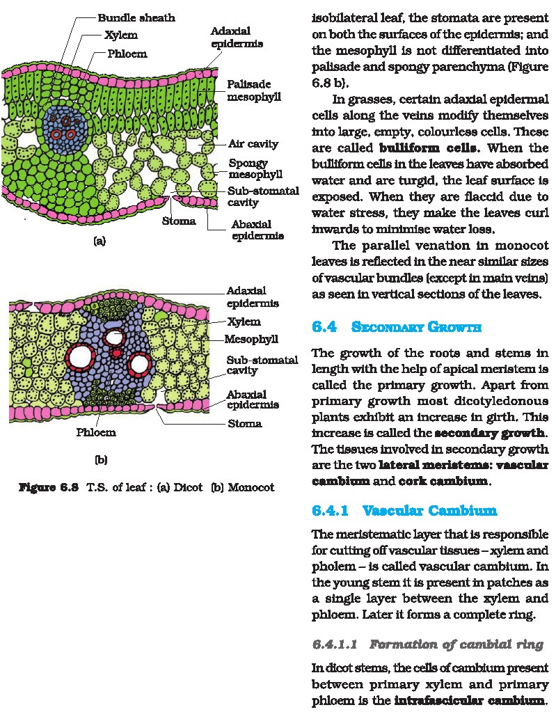
ANATOMY OF LEAF:Normally when we talk of a leaf, we
mean a foliage leaf is characterized by green colour, thinness and flatness.
The foliage leaves are lateral
appendages of a limited growth. They are very
important vegetative organs and chiefly concerned with the physiological
process like photosynthesis and transpiration.On the basis of mesophyll the
foliage are two types:
1. DORSIVENTRAL LEAF
2. ISOBILATERAL LEAF
|. lIsobilateral Leaf:
Mesophyll tissue is not differentiated and having same type of cells on both
adaxial and abaxial surfaces. These are commonly found in onocotyledonous
plants.
li. Dorsiventral Leaf:
In this the mesophyll tissues are differentiated into
palisade parenchyma and spongy parenchyma. It is
very common in the Dicotyledonous plants.
ANATOMY OF MONOCOT LEAF
(i) Epidermis:
a. Monocot leaf has upper and lower epidermis. =2.02:2
b. Cuticle is present on the outer wall stomata are
present on both upper and lower epidermis. Some cells of upper epidermis are
large and thin walled they are known
AS ote es,
(ii) Mesophyll:
a. Itis the ground tissue that is present between
both epidermal layers.
b. Mesophyll is not differentiated into palisade and
spongy parenchyma. The cells
are irregularly arranged with inter-cellular spaces.
(iii) Vascular bundles:
a. Large number of vascular bundles are present,
some of which are small and
some are large.
b.Vascular bundles are conjoint, collateral and
closed. Xylem is present towards
upper epidermis and phloem towards lower epidermis.
Phloem consists sieve tubes and companion cells. Xylem is formed of vessels,
tracheids and xylem parenchyma.
In Maize leaf the undifferentiated mesophyll occurs
in concentric layers
around vascular bundles having large centrifugal
chloroplasts in its large
bundle sheath cells. Such an arrangement is called .
ANATOMY OF DICOT LEAF
(i) Epidermis:
a. A dicotyledonous leaf is usually .
b. Ithas upper epidermis and lower epidermis. The
epidermis is generally formed of a single layer of cells which are packed
closely.
c. The cuticle on the upper epidermis is thicker as
compared to the lower
epidermis.
d. The minute openings that found on the epidermis
are termed as stomata. The
numbers of stomata are more on the upper surface
epidermis as compared to the
lower epidermis.
(ii) Mesophyll:
a. The entire tissue among the upper and lower
epidermis is termed as the
mesophyll.
b. In the mesophyll there are 2 regions. They are
spongy parenchyma and
palisade parenchyma.
c. Palisade parenchyma cells are observed below the
upper epidermis. It contains
vertically elongated cylindrical cells in one or
more than one layers. These cells are efficiently arranged with no
intercellular spaces. The task of palisade parenchyma is photosynthesis.
d. Spongy parenchyma takes place below the palisade
parenchyma. Spongy cells
are unevenly shaped. These cells are arranged very
loosely along with several
airspaces.
e. The air space which is found next to the stoma is
termed as respiratory cavity.
(iv) Vascular tissues:
a. These exist in the veins of leaf. VASCULAR
BUNDLES are collateral ,conjoint.
b. Xylem exists in the direction of the upper
epidermis; where else the phloem is in the direction of the lower epidermis.
c.termed as Bundle sheath. Phloem contains sieve
tubes, phloem parenchyma and companion cells. Phloem fibers are not present.
Xyiem contains vessels and
xylem parenchyma. Tracheids and xylem fibers are not
present.
PART: A_VERY SHORT ANSWER TYPE
QUESTIONS:
|. MULTIPLE CHOICE QUESTIONS:
1. Dicotyledonous
plans have:
a) Palisade parenchyma
b) Spongy parenchyma
c) Both (a) & (b)
d) None of the above
2. Leaves are:
a) Vegetative organs
b) Reproductive organs
c) Both (a) & (b)
d) None of the above
3. Vascular
Bundles in monocot leaf are:
a) Conjoint and closed
b) Conjoint and open
c) Radial and closed
d) None of the above
4. The epidermis
which covers upper surface of leaf is called:
a) Abascial epidermis
b) Adaxial epidermis
c) Both (a) & (b)
d) None of the above
5.
Monocotyledonous leaves are:
a) lsobilateral
b) Dorsiventral
c) Both (a) & (b)
d) None of the above
ll. Fill in the blanks:
1. Number of stomata is on the upper epidermis.
2. Dicot leaf show and type of vascular bundle.
3. cells are present in the upper epidermis of
monocot leaf.
III. State True or False:
1. Mesophyll in dicot leaf is differentiated into
palisade and spongy
parenchyma.
2. Spongy parenchyma consists of loosely arranged
irregularly shaped
cells.
ANSWER KEY: PART-A
l. MULTIPLE CHOICE QUESTIONS:
1. c- Mesophyll have both spongy and palisade
parenchyma.
2. a — Vegetative parts of plants.
3. a— Vascular bundle are conjoint and closed type.
4. b —-Adaxial epidermis
5. c - lsobilateral
ll. Fill in the blanks:
1. more
2. conjoint and collateral
3. Bulliform
Ill. State True or False:
1. True
2. True
PART: B SHORT ANSWER TYPE QUESTIONS:
1. Give difference between dicot and monocot leaf?
2. Briefly describe the vascular bundles in monocot
leaf?
PART: C LONG ANSWER TYPE QUESTIONS:
1. Draw a well labeled diagram of V.S. of dicot
leaf.
A70
INTRODUCTION:The
increase in thickness or girth due to the activity of cambium and
the cork cambium is known as secondary
growth.Secondary growth results from the activity of two meristem tissue
1. vascular cambium
2. Cork cambium: It occurs in the root and stem of gymnosperms and dicots.
Secondary growth in
dicot stem completes in following steps:
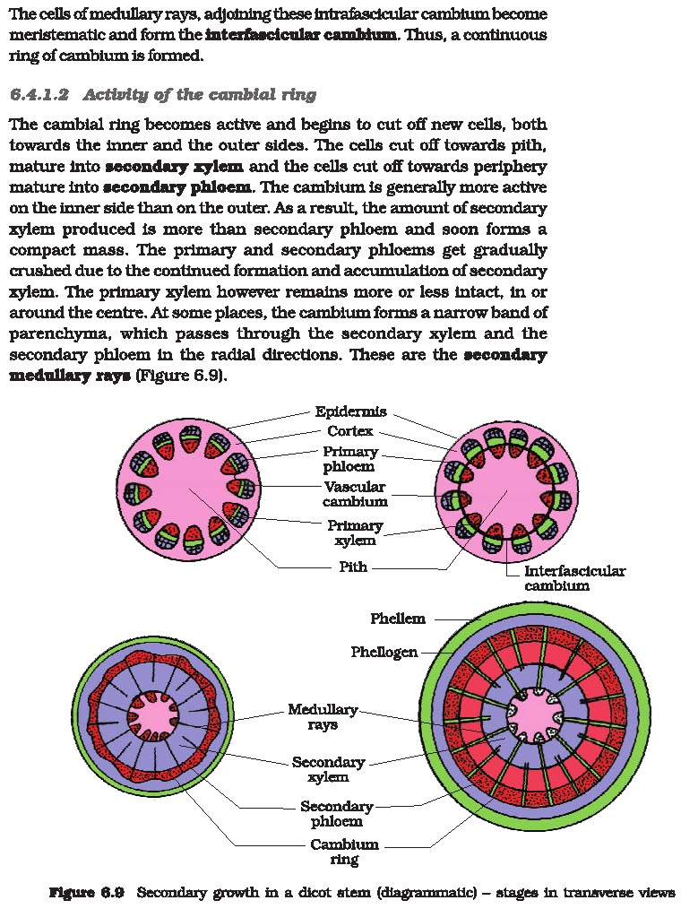
1. Formation of vascular cambium ring:
(i)Intrafascicular cambium:
It is primary in origin, present in between primary phloem and primary xylem.
At the time of secondary growth, it becomes functionally a secondary meristem
(ii) Interfascicular cambium.:it
is true secondary meristem. It originates from parenchyma cells of medullary
rays region. It lies in between the vascular bundles.
(iii) Vascular cambium ring: Both intrafascicular and interfascicular cambia join each other and forms cambium ring.
The cambial ring becomes active and begins to cut off new cells,
both towards the inner and the outer sides.The cells cut off towards pith,
mature into secondary xylem and the cells cut off towards periphery mature into
secondary phloem The cambium is generally more active on the inner side than on
the outer.As a result, the amount of secondary xylem produced is more than
secondary phloem and soon forms a compact mass.The primary and secondary
phloems get gradually crushed due to the continued formation and accumulation
of secondary xylem.The primary xylem however remains more or less intact, in or
around the centre.At some places, the cambium forms a narrow band of
parenchyma,
which passes through the secondary xylem and the secondary phloem in the radial directions. These are the secondary medullary rays.
These are formed by the seasonal activity of vascular cambium.
Cambium is not uniformly active throughout the
year.In spring or summer cambium is more active and forms large sized xylem
elements (vessels) which constitute spring or early wood.in autumn or winter,
cambium is less active and cut off small sized
xylem elements (vessels) and constitute autumn wood
or late wood.Both autumn and spring wood constitute an annual ring.
In one year, only one annual ring is formed.In
successive years numerous growth rings are formed.Thus, by counting the number
of annual rings in the main stem at the base we can determine the age of a
tree.This branch of science is known as dendrochronology.Growth rings are
distinct or sharply demarcated in the plants of
temperate climate, eg. Shimla, Nainital, Mussourie
due to presence of contrasting seasonal variations.Growth rings are not
distinct or sharply demarcated in the trees of tropical climate (near equator),
e.g. Calcutta, Bombay, Madras due to
absence of contrasting seasonal variations.
Formation of cork cambium:
1. It is formed by dedifferentiation of cortical
cells. It is also called as
phellogen. It is made of narrow thin-walled and
nearly rectangular cells.
2. Phellogen cuts cells on both sides. Towards inner
side, it forms
Rn Which is living and paranchymatous.
Towards outer side, it forms cork or phellem. It is
impervious to water.
3. Phellem + Phellogen + Phelloderm = Periderm
(Outer protective secondary tissue).
BARK OF TREE
BARK: It is a non-technical term that refers
to all tissues lying outside the
vascular cambium.It includes periderm and secondary
phloem.
(a) Bark formed early in the season is called early
or soft bark.
(b) Towards the end of the season late or hard bark
is formed
LENTICELS:
At some places phellogen cuts off closely arranged parenchymatous cells on the
outer side instead of cork cells. These parenchymatous cells soon rupture the
epidermis forming lens-shape opening called lenticels. These parenchymatous
cells are called complimentary cells.
BARK: SHOWING
LENTICELS It helps in exchange of gases.It is generally present in woody
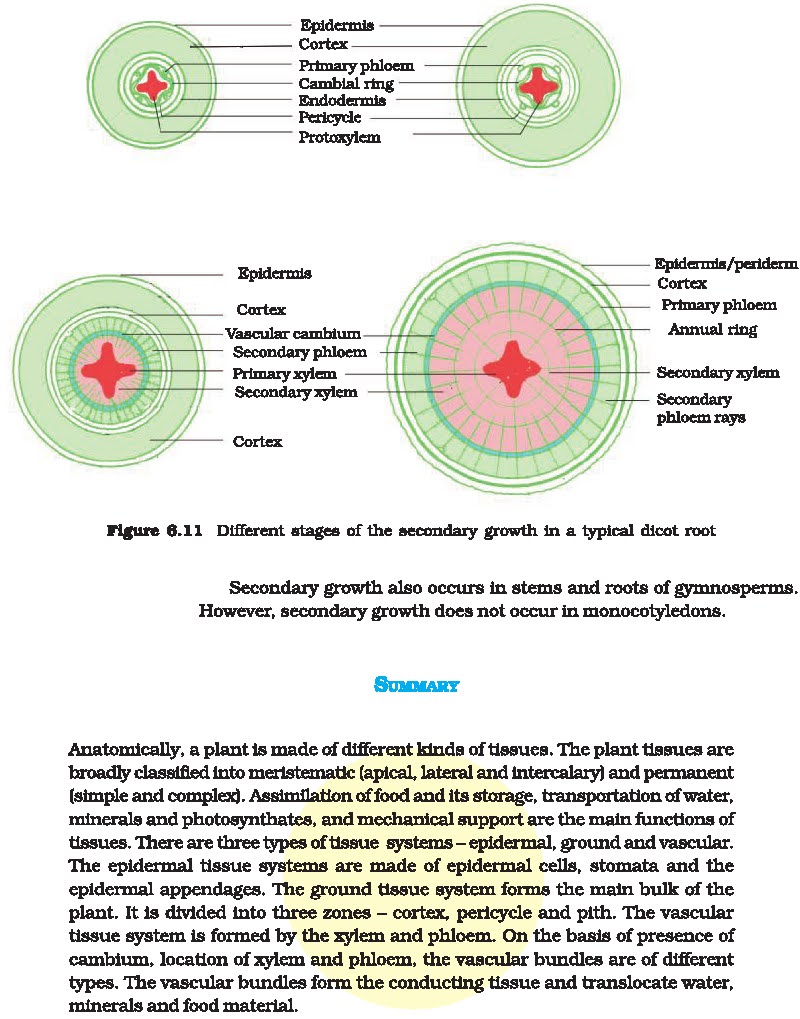
stem.Vascular bundles in dicot root are radial, exarch and mostly triarch.
Vascular cambium is formed secondarily from
conjuctive parenchyma cells lying just below each phioem strand. Thus, the
number of cambium strips formed equals the number of phloem strands.The cells
of pericycle lying outside the protoxylem also become
meristematic to form part of strips of cambium.These
cambial strips join the first formed cambium strips to form complete but wavy
ring of vascular cambium.This cambium ring produces secondary xylem on inner
side and secondary phloem on outer side.In roots, the growth rings are not
distinct because there is no seasonal variation under the soil.From the outer
layers of pericycle arises the phellogen which cuts
phellem (cork) on the outerside and secondary cortex
or phelloderm
toward the inner side
LET US KNOW WHAT WE HAVE LEARNT!
PART-A VERY SHORT ANSWER TYPE QUESTIONS:
A. MCQs:
1. Secondary
medullary rays are produced by:
(a) Fusiform initial
(b) Interfasicular cambium
(c) Phellogen
(d) Ray Initial
2. What is the
position of oldest secondary phloem:
(a) Just outside the pericycle
(b) Just outside the vascular cambium
(c) Just inside the pericycle
(d) Below the vascular cambium
3. Heart wood is
(a) Situated away from vascular cambium
(b) Situated near pith
(c) Nonfunctional
(d) All of these
4. In cork are
meant for gaseous exchange.
(a) Stomata
(b) Lenticels
(c) Bark
(d) Hydathodes
5. Cork cambium
is:
(a) Periderm
(b) Phellem
(c) Phelloderm
(d) Phellogen
B. FILL IN THE BLANKS:
1. Secondary Phloem is formed by .
2. In Leaf Anatomy, Phloem is directed towards .
3. Heartwoodis sin color.
C. TRUE/FALSE:
1. Heart wood is more hard durable and resistant to
the attack of micro-
organisms and insects.
2. Cork Cambium is called Complimentary Cells.
ANSWER KEY: PART -A
A. MCQs:
1.(b) Interfasicular cambium(They are formed by
activity of fasicular
cambium)
2. (b) Just outside the vascular cambium. (the
position of oldest secondary
phloem is Just outside the vascular cambium)
3. (b) Situated near pith. (Heart wood isSituated
near pith& forms the
central major portion)
4. (b) Lenticels(In cork Lenticelsare meant for
gaseous exchange)
5. (d) Phellogen(Cork cambium is also called as
Phellogen)
B. FILL Ups:
1. vascular cambium
2. lower surface
3. brown
C. TRUE/FALSE:
1. True
2. False (Cork cambium is also called as Phellogen)
PART -B SHORT ANSWER TYPE QUESTIONS
1. How annual rings are formed?
2. How is cambial ring formed in dicot stem?
PART-C LONG ANSWER TYPE QUESTIONS
1. Describe the process of secondary growth in Dicot
Stem.
A71
RECAPITULATION:Dear
students in the chapter ‘Anatomy of flowering plants ‘.you have read all the
topics in detail. As this chapter deals with the internal structure of plants,
We have learnt that Plants have cells as the basic unit, cells are organized
into tissues and in turn the tissues are organized into organs. Different
organs in a plant show
difference in their structure. Now let us do NCERT
book questions;
2. Cork cambium forms
tissues that form the cork. Do you agree with this statement? Explain.
ANS. Yes, cork cambium forms tissues that in turn
form the [J When the stem
increases in girth, another meristematic tissue
known as phellogen or cork
cambium grows in the cortex region of the stem. This
phellogen cuts off cells on
both of the sides. The outer cells differentiate
into the [MM or the cork while the inner cells differentiate into the EM or
secondary cortex. The cork is
impermeable to water because of suberin, rendering
protection to the tissues
undemeath.
3. Explain the
process of secondary growth in the stems of woody angiosperms with the help of
schematic diagrams. What is its significance?
ANS. The primary xylem and phloem exhibits the
presence of a strip of cambium
in woody dicot roots known as the interfascicular cambium which is formed from the cells of the medullary rays connecting the interfascicular cambium. Hence, the continuous cambium ring is formed. The cambium separates from the newly formed celis on either side while the cells found towards the exterior differentiate into the secondary phloem. The cells detach towards the pith giving rise to the secondary xylem. The secondary xylem is synthesized in excess compared to the secondary phloem. When there is secondary growth in plants, the girth of the
plants increase, along with an
increase in the water content and nutrients in order
to assist the ever growing leaves, rendering support
to the plants.
6. The transverse
section of a plant material shows the following anatomical
features (a) the vascular bundles are conjoint,
scattered and surrounded by sclerenchymatous bundle sheaths.
(b) phloem parenchyma is absent. What will you
identify it as?
ANS. The transverse section is of Monocot stem. It
is because the vascular
bundles are dispersed in monocot stems. The phloem
parenchyma is not found.
7. Why are xylem
and phloem called complex tissues?
ANS. Xylem and Phioem are called complex tissues
because they are made of more than one type of cells which work together as a
unit to perform the function.Xylem transports water while phloem transports
food.
8. What is a
stomatal apparatus? Explain the structure of stomata with a
labelled diagram.
ANS. Stomata are structures present in the epidermis
of leaves. Stomata regulate
the process of transpiration and gaseous exchange.
Each stoma is composed of
two bean-shaped cells known as guard cells which enclose stomatal pore. Guard cells are dumbbell-shaped, where its outer wall is thin and inner wall is highly thickened. These structures possess chloroplasts and regulate the closing
and opening of the stomata. The epidermal cells near
the guard cells in some cases become specialized in their structure, shape and
size; they are referred to as subsidiary cells. The guard-cells, the stomatal
aperture and girdling subsidiary cells are collectively referred toas stomatal
apparatus.
9. Name the three
basic tissue systems in the flowering plants. Give the tissue names under each
system.
ANS. Following are the three basic tissue systems in
the flowering plants.Epidermal tissue system Epidermal tissue system includes
epidermis and epidermal appendages.Epidermis comprises epidermal cells and
guard cells while the epidermal
appendages include root hair, stem hair, stinging hair
and glandular hair.The qround tissue system Ground tissue system is made up of
simple tissues such as parenchyma,
collenchyma and sclerenchyma.Vascular tissue system
Vascular tissue system consists of complex tissues such as Xylem, phloem and
vascular cambium.
10. How is the
study of plant anatomy useful to us?
ANS. ie BEE Physiological conditions can be studied,
which helps in crop improvement. Study of plant fibres such as flax, jute etc.
helps in their commercial
exploitation as it enables them to predict the
strength of wood which can be utilized
to its potential.
11. What is
periderm? How does periderm formation take place in the dicot
stems?
ANS. Phetieges; EE and phelloderm are collectively
known as periderm.While plants undergo secondary growth, the outer epidermal
layer and the cortical layer are ripped due to cambium. In order to replace
them, the cortex ceils tum meristematic which produces the cork cambium or the
phellogen which comprises thin-walled, narrow and rectangular cells. The phellogen
sheds cells on either side.cork The cells which shed from the exterior give
rise to the or phellem. The suberin accumulates in its cell wail making it
impermeable to water while the inner cellsemerge to become the secondary cortex
or phelloderm which is parenchymatous.
12. Describe the
internal structure of a dorsiventral leaf with the help of
labelled diagram.
ANS. Dicots exhibit dorsiventral leaves. When
examined, the vertical section
of a dorsiventral leaf consists of three different
parts, they are:
Epidermis —
it is found on the adaxial epidermis (upper surface) and the abaxial epidermis
(lower surface). On the outside, the epidermis is covered with a thick cuticle.
Compared to the upper surface, the abaxial epidermis comprises more stomata.
Mesophylil -
it is a tissue found in between the abaxial and adaxial : epidermises. This
tissue is differentiated into the palisade parenchyma and the spongy
parenchyma. The palisade parenchyma is composed of tall,compactly-arranged
cells while the spongy parenchyma comprises round or oval, loosely-arranged
cells possessing intercellular spaces. The mesophyll comprises chloroplasts
that carry out photosynthesis.
Vascular system
— The vascular bundles that are found in leaves are closed and conjoint which
are engirdied by thick layers of bundle-sheath cells.
A72
RECAPITULATION:Dear students,
earlier you have studied Anatomy of flowering plants. Today we will revise all
important diagrams from chapter 6 — Anatomy of flowering plants.
THE TISSUE:-A
Tissue is a group of cells having a common origin and usually performing common
function.
TYPES OF PLANT TISSUE
1. MERISTEMATIC TISSUE:-
Cells are capable of cell division.
APICAL MERISTEM:
- FOUND AT THE TIPS ON THE SHOOT AND ROOTS.
2. PERMANENT TISSUE: -
Incapable of cell division, long lasting tissue which have mature cell.
SIMPLE TISSUE:
- Made of only one type of cells.
COMPLEX TISSUE:
- Made of more than one type of cells.
PART -A VERY SHORT ANSWER TYPE QUESTIONS:
1. The most
common ground tissue in plant is:
a) Epidermis
b) Cortex
c) Parenchyma
d) Collenchymas
2. The outer wall
of epidermal cells contains a fatty substance called:
a) Cutin
b) Melanin
c) Cuticle
d) None of these
3. Vascular
tissue system consists of
a) Xylem
b) Phloem
c) Sclerenchyma
d) Both a and b
4.Root cap in
dicots is formed from
a) Ground meristem
b) Procambium
c) Protoderm
d) Calyptrogens
5.Each vascular
bundle is covered by sclerenchyma bundle sheath in
a) Monocot stem
b) Dicot stem
c) Both
d) None
B.Fill in the blanks:
1. Internal plant organisation is studied under .
2. Dicot leaf show and type of vascular bundles.
3. is regarded as father of plant anantomy.
C. TRUE/FALSE
1.Study of tissue is called HISTOGRAM.
2. Parenchyma ,collenchyma,sclerenchyma form
vascular tissue system.
3.Roots cap is cushion of parenchymatous tissue for
protection.
ANSWER KEY: PART -A
MCQs
1. (c) Parenchyma most common ground tissue.
2. (c) Cutin outer wall of epidermal cells contain
fatty substances.
3. (d) sclerenchyma is a ground tissue.
4. (c) In dicots the root cap is derived from the
protoderm.
5. (b) In dicots vascular bundles are covered by
sclerenchymatous bundle sheath.
B.Fill in the blanks:
1. Anatomy
2. Conjoint and Collateral
3. N. Grew
C. TRUE/FALSE
1. False (study of tissue is called HISTOLOGY.
2. False (xylem and phloem form vascular tissue
system).
3. True
PART-B SHORT ANSWER TYPE QUESTIONS
1. Name the component of xylem which of these is
suitable for carrying
water?
2. What is the function of sclerenchyma cells?
3. Give the differences between Dicot and Monocot
roots.
PART-C LONG ANSWER TYPE QUESTIONS
1. Draw a well labelled diagram of stomata which
shows opening and closing
of stomata. Also write the main functions of
stomata.
2. Draw a well labelled diagram of a typical monocot
root and explain its
different parts.


